 Movie
Movie Controller


+ Open data

- Basic information
| Entry | Database: EMDB / ID: EMD-4319 | |||||||||
|---|---|---|---|---|---|---|---|---|---|---|
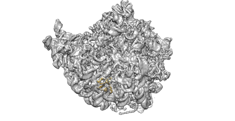
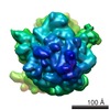
| Title | uL23 beta hairpin loop deletion of E.coli ribosome | |||||||||
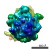
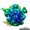
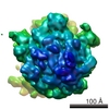
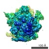
 Map data Map data | Sharpened map of E.coli ribosome with the beta-hairpin of uL23 deleted. | |||||||||
 Sample Sample |
| |||||||||
 Keywords Keywords | uL23 / loop deletion / ribosomal tunnel / RIBOSOMAL PROTEIN | |||||||||
| Function / homology |  Function and homology information Function and homology informationribosomal large subunit assembly / cytosolic large ribosomal subunit / cytoplasmic translation / rRNA binding / structural constituent of ribosome / ribosome / translation / ribonucleoprotein complex / cytoplasm Similarity search - Function | |||||||||
| Biological species |  | |||||||||
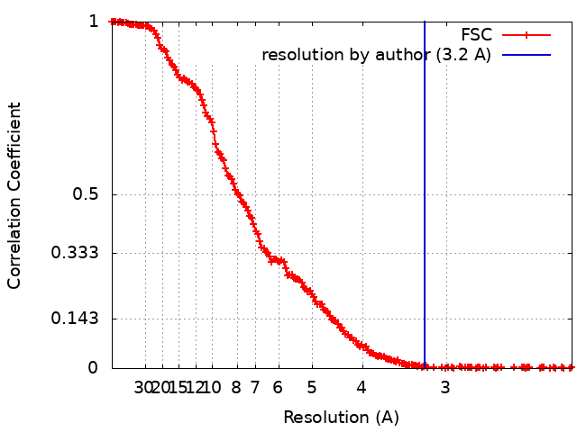
| Method | single particle reconstruction / cryo EM / Resolution: 3.2 Å | |||||||||
 Authors Authors | Kudva R / von Heijne G | |||||||||
| Funding support |  Sweden, 1 items Sweden, 1 items
| |||||||||
 Citation Citation | Journal: Elife / Year: 2018 Title: The shape of the bacterial ribosome exit tunnel affects cotranslational protein folding. Authors: Renuka Kudva / Pengfei Tian / Fátima Pardo-Avila / Marta Carroni / Robert B Best / Harris D Bernstein / Gunnar von Heijne /  Abstract: The ribosome exit tunnel can accommodate small folded proteins, while larger ones fold outside. It remains unclear, however, to what extent the geometry of the tunnel influences protein folding. ...The ribosome exit tunnel can accommodate small folded proteins, while larger ones fold outside. It remains unclear, however, to what extent the geometry of the tunnel influences protein folding. Here, using ribosomes with deletions in loops in proteins uL23 and uL24 that protrude into the tunnel, we investigate how tunnel geometry determines where proteins of different sizes fold. We find that a 29-residue zinc-finger domain normally folding close to the uL23 loop folds deeper in the tunnel in uL23 Δloop ribosomes, while two ~ 100 residue proteins normally folding close to the uL24 loop near the tunnel exit port fold at deeper locations in uL24 Δloop ribosomes, in good agreement with results obtained by coarse-grained molecular dynamics simulations. This supports the idea that cotranslational folding commences once a protein domain reaches a location in the exit tunnel where there is sufficient space to house the folded structure. | |||||||||
| History |
|
- Structure visualization
| Movie |
Movie viewer |
|---|---|
| Structure viewer | EM map: SurfViewMolmilJmol/JSmol |
| Supplemental images |
- Downloads & links
-EMDB archive
| Map data | emd_4319.map.gz | 347.4 MB | EMDB map data format | |
|---|---|---|---|---|
| Header (meta data) | emd-4319-v30.xmlemd-4319.xml | 16.1 KB 16.1 KB | Display Display | EMDB header |
| FSC (resolution estimation) | emd_4319_fsc.xml | 19.1 KB | Display | FSC data file |
| Images |  emd_4319.png emd_4319.png | 172.3 KB | ||
| Masks | emd_4319_msk_1.map | 371.3 MB | Mask map | |
| Filedesc metadata | emd-4319.cif.gz | 5.6 KB | ||
| Others | emd_4319_half_map_1.map.gzemd_4319_half_map_2.map.gz | 344.3 MB 344.3 MB | ||
| Archive directory |  http://ftp.pdbj.org/pub/emdb/structures/EMD-4319ftp://ftp.pdbj.org/pub/emdb/structures/EMD-4319 http://ftp.pdbj.org/pub/emdb/structures/EMD-4319ftp://ftp.pdbj.org/pub/emdb/structures/EMD-4319 | HTTPS FTP |
-Related structure data
| Related structure data |  6fu8MC M: atomic model generated by this map C: citing same article ( |
|---|---|
| Similar structure data |


-Links
| EMDB pages | EMDB (EBI/PDBe) / EMDataResource |
|---|---|
| Related items in Molecule of the Month |


-Map
| File | Download / File: emd_4319.map.gz / Format: CCP4 / Size: 371.3 MB / Type: IMAGE STORED AS FLOATING POINT NUMBER (4 BYTES) | ||||||||||||||||||||||||||||||||||||||||||||||||||||||||||||
|---|---|---|---|---|---|---|---|---|---|---|---|---|---|---|---|---|---|---|---|---|---|---|---|---|---|---|---|---|---|---|---|---|---|---|---|---|---|---|---|---|---|---|---|---|---|---|---|---|---|---|---|---|---|---|---|---|---|---|---|---|---|
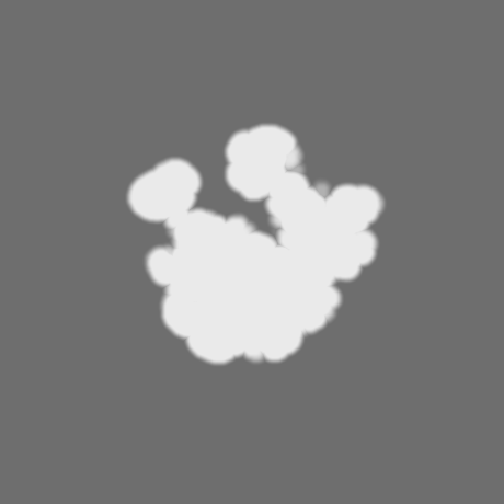
| Annotation | Sharpened map of E.coli ribosome with the beta-hairpin of uL23 deleted. | ||||||||||||||||||||||||||||||||||||||||||||||||||||||||||||
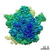
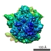
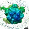
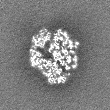
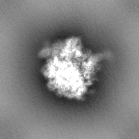
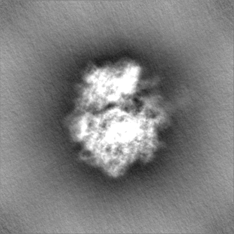
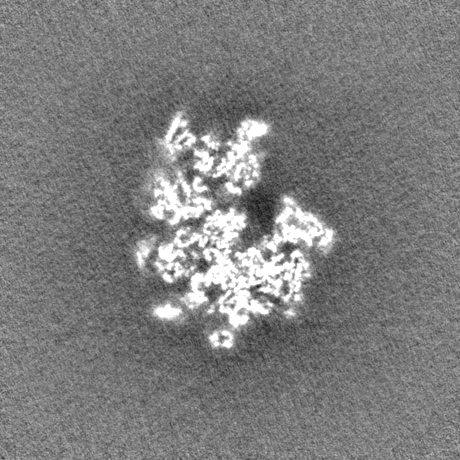
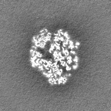
| Projections & slices | Image control
Images are generated by Spider. | ||||||||||||||||||||||||||||||||||||||||||||||||||||||||||||
| Voxel size | X=Y=Z: 1.09 Å | ||||||||||||||||||||||||||||||||||||||||||||||||||||||||||||
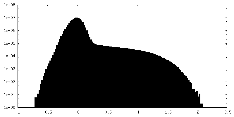
| Density |
| ||||||||||||||||||||||||||||||||||||||||||||||||||||||||||||
| Symmetry | Space group: 1 | ||||||||||||||||||||||||||||||||||||||||||||||||||||||||||||
| Details | EMDB XML:
CCP4 map header:
| ||||||||||||||||||||||||||||||||||||||||||||||||||||||||||||
 Z (Sec.)
 Y (Row.)
 X (Col.)


-Supplemental data
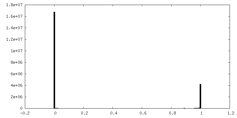
-Mask #1
| File | emd_4319_msk_1.map | ||||||||||||
|---|---|---|---|---|---|---|---|---|---|---|---|---|---|
| Projections & Slices |
| ||||||||||||
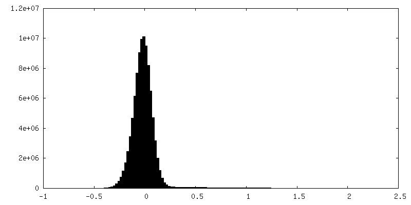
| Density Histograms |

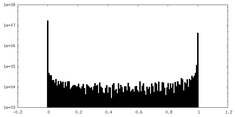
-Half map: Half map A of E.coli ribosome with the beta-hairpin of uL23 deleted.
| File | emd_4319_half_map_1.map | ||||||||||||
|---|---|---|---|---|---|---|---|---|---|---|---|---|---|
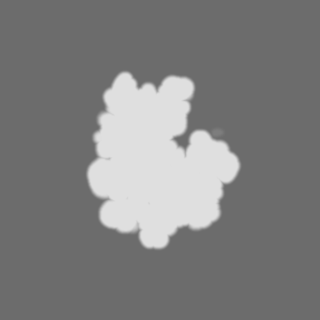
| Annotation | Half map A of E.coli ribosome with the beta-hairpin of uL23 deleted. | ||||||||||||
| Projections & Slices |
| ||||||||||||
| Density Histograms |


-Half map: Half map B of E.coli ribosome with the beta-hairpin of uL23 deleted.
| File | emd_4319_half_map_2.map | ||||||||||||
|---|---|---|---|---|---|---|---|---|---|---|---|---|---|
| Annotation | Half map B of E.coli ribosome with the beta-hairpin of uL23 deleted. | ||||||||||||
| Projections & Slices |
| ||||||||||||
| Density Histograms |


- Sample components
-Entire : E.coli ribosome with beta-hairpin of uL23 deleted
| Entire | Name: E.coli ribosome with beta-hairpin of uL23 deleted |
|---|---|
| Components |
|
-Supramolecule #1: E.coli ribosome with beta-hairpin of uL23 deleted
| Supramolecule | Name: E.coli ribosome with beta-hairpin of uL23 deleted / type: complex / ID: 1 / Parent: 0 / Macromolecule list: all Details: Deposited PDB is of the uL23 with the beta-hairpin loop deleted. |
|---|---|
| Source (natural) | Organism: |
| Molecular weight | Theoretical: 2.6 MDa |
-Macromolecule #1: 50S ribosomal protein L23
| Macromolecule | Name: 50S ribosomal protein L23 / type: protein_or_peptide / ID: 1 / Number of copies: 1 / Enantiomer: LEVO |
|---|---|
| Source (natural) | Organism: |
| Molecular weight | Theoretical: 9.324003 KDa |
| Recombinant expression | Organism: |
| Sequence | String: MIREERLLKV LRAPHVSEKA STAMEKSNTI VLKVAKDATK AEIKAAVQKL FEVEVEVVNT LVVKRRSDWK KAYVTLKEGQ NL UniProtKB: Large ribosomal subunit protein uL23 |
-Experimental details
-Structure determination
| Method | cryo EM |
|---|---|
 Processing Processing | single particle reconstruction |
| Aggregation state | particle |
-Sample preparation
| Buffer | pH: 7.5 |
|---|---|
| Grid | Material: COPPER / Mesh: 400 / Support film - Material: CARBON / Support film - topology: LACEY / Support film - Film thickness: 0.02 / Pretreatment - Type: GLOW DISCHARGE / Pretreatment - Time: 40 sec. / Pretreatment - Atmosphere: AIR / Pretreatment - Pressure: 0.0003 kPa |
| Vitrification | Cryogen name: ETHANE / Chamber humidity: 100 % / Chamber temperature: 277 K / Instrument: FEI VITROBOT MARK IV |
- Electron microscopy
| Microscope | FEI TITAN KRIOS |
|---|---|
| Image recording | Film or detector model: FEI FALCON II (4k x 4k) / Detector mode: INTEGRATING / Number grids imaged: 1 / Number real images: 3500 / Average electron dose: 1.17 e/Å2 |
| Electron beam | Acceleration voltage: 300 kV / Electron source:  FIELD EMISSION GUN FIELD EMISSION GUN |
| Electron optics | C2 aperture diameter: 70.0 µm / Illumination mode: FLOOD BEAM / Imaging mode: BRIGHT FIELD / Cs: 2.7 mm / Nominal defocus max: 3.0 µm / Nominal defocus min: 1.0 µm |
| Sample stage | Specimen holder model: FEI TITAN KRIOS AUTOGRID HOLDER / Cooling holder cryogen: NITROGEN |
| Experimental equipment |  Model: Titan Krios / Image courtesy: FEI Company |
+Image processing
-Atomic model buiding 1
| Refinement | Protocol: RIGID BODY FIT |
|---|---|
| Output model | PDB-6fu8: |
