 Movie
Movie Controller

[English] 日本語
 Yorodumi
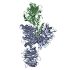
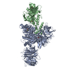
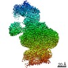
- PDB-4e5z: Damaged DNA induced UV-damaged DNA-binding protein (UV-DDB) dimer... -
+ Open data

- Basic information
| Entry | Database: PDB / ID: 4e5z | ||||||
|---|---|---|---|---|---|---|---|
| Title | Damaged DNA induced UV-damaged DNA-binding protein (UV-DDB) dimerization and its roles in chromatinized DNA repair | ||||||
 Components Components |
| ||||||
 Keywords Keywords | DNA BINDING PROTEIN/DNA / BETA BARREL / PROTEIN-DNA COMPLEX / DOUBLE HELIX / damage / DNA repair / Host-virus interactions / Protein ubiquitination / Proteosomal degradation / DNA BINDING PROTEIN-DNA complex | ||||||
| Function / homology |  Function and homology information Function and homology informationpositive regulation by virus of viral protein levels in host cell / spindle assembly involved in female meiosis / epigenetic programming in the zygotic pronuclei / UV-damage excision repair / biological process involved in interaction with symbiont / regulation of mitotic cell cycle phase transition / WD40-repeat domain binding / Cul4A-RING E3 ubiquitin ligase complex / Cul4-RING E3 ubiquitin ligase complex / Cul4B-RING E3 ubiquitin ligase complex ...positive regulation by virus of viral protein levels in host cell / spindle assembly involved in female meiosis / epigenetic programming in the zygotic pronuclei / UV-damage excision repair / biological process involved in interaction with symbiont / regulation of mitotic cell cycle phase transition / WD40-repeat domain binding / Cul4A-RING E3 ubiquitin ligase complex / Cul4-RING E3 ubiquitin ligase complex / Cul4B-RING E3 ubiquitin ligase complex / ubiquitin ligase complex scaffold activity / negative regulation of reproductive process / negative regulation of developmental process / pyrimidine dimer repair / viral release from host cell / cullin family protein binding / ectopic germ cell programmed cell death / response to UV / positive regulation of viral genome replication / protein autoubiquitination / site of DNA damage / proteasomal protein catabolic process / positive regulation of gluconeogenesis / TP53 Regulates Transcription of DNA Repair Genes / nucleotide-excision repair / sperm end piece / regulation of circadian rhythm / Recognition of DNA damage by PCNA-containing replication complex / DNA Damage Recognition in GG-NER / Dual Incision in GG-NER / Wnt signaling pathway / Transcription-Coupled Nucleotide Excision Repair (TC-NER) / Formation of TC-NER Pre-Incision Complex / Formation of Incision Complex in GG-NER / protein polyubiquitination / Dual incision in TC-NER / positive regulation of protein catabolic process / Gap-filling DNA repair synthesis and ligation in TC-NER / cellular response to UV / cell junction / rhythmic process / site of double-strand break / sperm principal piece / Neddylation / sperm midpiece / ubiquitin-dependent protein catabolic process / damaged DNA binding / proteasome-mediated ubiquitin-dependent protein catabolic process / protein-macromolecule adaptor activity / chromosome, telomeric region / Ub-specific processing proteases / protein ubiquitination / DNA repair / apoptotic process / DNA damage response / negative regulation of apoptotic process / chromatin / protein-containing complex binding / nucleolus / protein-containing complex / : / DNA binding / extracellular exosome / nucleoplasm / nucleus / cytoplasm Similarity search - Function | ||||||
| Biological species |  Homo sapiens (human) Homo sapiens (human) | ||||||
| Method |  X-RAY DIFFRACTION / SYNCHROTRON / MOLECULAR REPLACEMENT / Resolution: 3.22 Å X-RAY DIFFRACTION / SYNCHROTRON / MOLECULAR REPLACEMENT / Resolution: 3.22 Å | ||||||
 Authors Authors | Yeh, J.I. / Du, S. | ||||||
 Citation Citation | Journal: Proc.Natl.Acad.Sci.USA / Year: 2012 Title: Damaged DNA induced UV-damaged DNA-binding protein (UV-DDB) dimerization and its roles in chromatinized DNA repair. Authors: Yeh, J.I. / Levine, A.S. / Du, S. / Chinte, U. / Ghodke, H. / Wang, H. / Shi, H. / Hsieh, C.L. / Conway, J.F. / Van Houten, B. / Rapic-Otrin, V. | ||||||
| History |
|
- Structure visualization
| Structure viewer | Molecule: MolmilJmol/JSmol |
|---|
- Downloads & links
-Download
| PDBx/mmCIF format | 4e5z.cif.gz | 666.9 KB | Display | PDBx/mmCIF format |
|---|---|---|---|---|
| PDB format | pdb4e5z.ent.gz | 539 KB | Display | PDB format |
| PDBx/mmJSON format | 4e5z.json.gz | Tree view | PDBx/mmJSON format | |
| Others |  Other downloads Other downloads |
-Validation report
| Arichive directory | https://data.pdbj.org/pub/pdb/validation_reports/e5/4e5zftp://data.pdbj.org/pub/pdb/validation_reports/e5/4e5z | HTTPS FTP |
|---|
-Related structure data
| Related structure data | 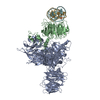 4e54C 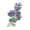 3ei2S C: citing same article ( S: Starting model for refinement |
|---|---|
| Similar structure data |


-Links
 PDBj
PDBj


- Assembly
| Deposited unit | 
| ||||||||
|---|---|---|---|---|---|---|---|---|---|
| 1 |
| ||||||||
| Unit cell |
|
-Components
| #1: Protein | Mass: 128478.914 Da / Num. of mol.: 1 Source method: isolated from a genetically manipulated source Source: (gene. exp.) Homo sapiens (human) / Gene: DDB1, DDB1_HUMAN, Q16531, XAP1Plasmid: pBlueBac4.5/V5-His NT-His10-DDB1pBlueBac4.5/V5-His NT-His10-DDB1 Cell line (production host): Sf9 / Production host:   Spodoptera frugiperda (fall armyworm) / References: UniProt: Q16531 Spodoptera frugiperda (fall armyworm) / References: UniProt: Q16531 |
|---|---|
| #2: Protein | Mass: 49059.004 Da / Num. of mol.: 1 Source method: isolated from a genetically manipulated source Source: (gene. exp.) Homo sapiens (human) / Gene: DDB2 / Plasmid: pBlueBac4.5/V5-HisNT-FLAG-DDB2 / Cell line (production host): Sf9 / Production host: Spodoptera frugiperda (fall armyworm) / References: UniProt: Q92466 |
| #3: DNA chain | Mass: 7424.801 Da / Num. of mol.: 1 / Source method: obtained synthetically Details: Synthetic single stranded 24-oligodeoxynucleotides with complementary strand sequence: 5-TGACTGTATGATGACGATGCTGAC-3 |
| #4: DNA chain | Mass: 7189.646 Da / Num. of mol.: 1 / Source method: obtained synthetically Details: Synthetic single stranded oligodeoxynucleotides with a central tetrahydrofuran abasic site mimic (3DR) on coding strand with sequence: 5-GTCAGCATCG(3DR)CATCATACAGTCA-3 |
| Has protein modification | Y |
-Experimental details
-Experiment
| Experiment | Method: X-RAY DIFFRACTION / Number of used crystals: 3 |
|---|
- Sample preparation
| Crystal | Density Matthews: 2.81 Å3/Da / Density % sol: 56.19 % |
|---|---|
| Crystal grow | Temperature: 277 K / Method: vapor diffusion / pH: 7.5 Details: 20mM Tris pH 7.5, 2mM MgCl2, 1mM EDTA, 2mM TECP, 5% Glycerol, 0.02% azide. UV-DDB-AP24 complex (molar ratio of 1:3 UV-DDB:DNA) at 2.5 mg/mL. 'AP24' refers to synthetic DNA substrate of 24- ...Details: 20mM Tris pH 7.5, 2mM MgCl2, 1mM EDTA, 2mM TECP, 5% Glycerol, 0.02% azide. UV-DDB-AP24 complex (molar ratio of 1:3 UV-DDB:DNA) at 2.5 mg/mL. 'AP24' refers to synthetic DNA substrate of 24-bpr with a central abasic site mimic., VAPOR DIFFUSION, temperature 277K |
-Data collection
| Diffraction | Mean temperature: 100 K |
|---|---|
| Diffraction source | Source: SYNCHROTRON / Site: APS  / Beamline: 23-ID-D / Beamline: 23-ID-D |
| Detector | Type: MARMOSAIC 225 mm CCD / Detector: CCD / Date: Jul 8, 2009 / Details: monochromators |
| Radiation | Monochromator: SAGITALLY FOCUSED Si(111) / Protocol: SINGLE WAVELENGTH / Monochromatic (M) / Laue (L): M / Scattering type: x-ray |
| Radiation wavelength | Relative weight: 1 |
| Reflection | Resolution: 3.2→41.093 Å / Num. all: 36260 / Num. obs: 33928 / % possible obs: 77.8 % / Observed criterion σ(F): 2 / Observed criterion σ(I): 2 / Redundancy: 5.6 % / Biso Wilson estimate: 38.09 Å2 / Rmerge(I) obs: 0.117 / Rsym value: 0.105 / Net I/σ(I): 10.9 |
| Reflection shell | Resolution: 3.2→3.31 Å / Redundancy: 3.6 % / Rmerge(I) obs: 0.117 / Mean I/σ(I) obs: 3.1 / Rsym value: 0.358 / % possible all: 77.8 |
- Processing
| Software |
| ||||||||||||||||||||||||||||||||||||||||||||||||||||||||||||||||||||||||||||||||||||||||||||||||||||
|---|---|---|---|---|---|---|---|---|---|---|---|---|---|---|---|---|---|---|---|---|---|---|---|---|---|---|---|---|---|---|---|---|---|---|---|---|---|---|---|---|---|---|---|---|---|---|---|---|---|---|---|---|---|---|---|---|---|---|---|---|---|---|---|---|---|---|---|---|---|---|---|---|---|---|---|---|---|---|---|---|---|---|---|---|---|---|---|---|---|---|---|---|---|---|---|---|---|---|---|---|---|
| Refinement | Method to determine structure: MOLECULAR REPLACEMENT Starting model: 3EI2 Resolution: 3.22→41.093 Å / Cor.coef. Fo:Fc: 0.968 / Cor.coef. Fo:Fc free: 0.964 / SU ML: 0.44 / σ(F): 0 / Phase error: 30.96 / Stereochemistry target values: MLHL Details: THE MODEL WAS REFINED USING ITERATIVE CYCLES OF TLS AND RESTRAINED REFINEMENT (INCLUDING SECONDARY STRUCTURE, GEOMETRY, AND TORSION ANGLE RESTRAINTS) THROUGH PHENIX. CAREFUL INSPECTION OF ...Details: THE MODEL WAS REFINED USING ITERATIVE CYCLES OF TLS AND RESTRAINED REFINEMENT (INCLUDING SECONDARY STRUCTURE, GEOMETRY, AND TORSION ANGLE RESTRAINTS) THROUGH PHENIX. CAREFUL INSPECTION OF WEIGHTED AND UNWEIGHTED MAPS, IN PARTICULAR, THE DIFFERENCE FOURIER MAPS, AFTER EACH REFINEMENT ROUND VERIFIED CORRECTNESS OF REGIONS MODIFIED OR EXTENDED IN THE PREVIOUS CYCLE. PROGRAMMATIC DIFFERENCES IN THE APPLICATION AND SCALING OF TLS PARAMETERS MAY RESULT IN VARIATIONS IN THE MAPS CALCULATED USING THE SF DIRECTLY DOWNLOADED FROM THE DATABASE. CALCULATING STRUCTURE FACTORS (SF) USING MODEL COORDINATES AND THERMAL PARAMETERS FROM THE DEPOSITED PDB FILES IN PHENIX WILL REPRODUCE THE MAPS AND CONFORMATIONAL FEATURES DESCRIBED BY THE AUTHORS IN THE CITATION.
| ||||||||||||||||||||||||||||||||||||||||||||||||||||||||||||||||||||||||||||||||||||||||||||||||||||
| Solvent computation | Shrinkage radii: 0.9 Å / VDW probe radii: 1.11 Å / Solvent model: FLAT BULK SOLVENT MODEL | ||||||||||||||||||||||||||||||||||||||||||||||||||||||||||||||||||||||||||||||||||||||||||||||||||||
| Displacement parameters | Biso mean: 185.363 Å2
| ||||||||||||||||||||||||||||||||||||||||||||||||||||||||||||||||||||||||||||||||||||||||||||||||||||
| Refinement step | Cycle: LAST / Resolution: 3.22→41.093 Å
| ||||||||||||||||||||||||||||||||||||||||||||||||||||||||||||||||||||||||||||||||||||||||||||||||||||
| Refine LS restraints |
| ||||||||||||||||||||||||||||||||||||||||||||||||||||||||||||||||||||||||||||||||||||||||||||||||||||
| LS refinement shell |
| ||||||||||||||||||||||||||||||||||||||||||||||||||||||||||||||||||||||||||||||||||||||||||||||||||||
| Refinement TLS params. | Method: refined / Refine-ID: X-RAY DIFFRACTION
| ||||||||||||||||||||||||||||||||||||||||||||||||||||||||||||||||||||||||||||||||||||||||||||||||||||
| Refinement TLS group |
|
