 Movie
Movie Controller

[English] 日本語
 Yorodumi
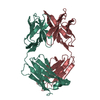
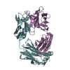
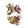
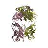
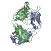
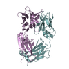
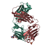
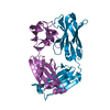
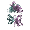
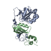
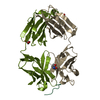
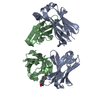
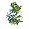
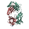
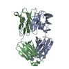
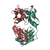
- PDB-8fab: CRYSTAL STRUCTURE OF THE FAB FRAGMENT FROM THE HUMAN MYELOMA IMMU... -
+ Open data

- Basic information
| Entry | Database: PDB / ID: 8fab | ||||||
|---|---|---|---|---|---|---|---|
| Title | CRYSTAL STRUCTURE OF THE FAB FRAGMENT FROM THE HUMAN MYELOMA IMMUNOGLOBULIN IGG HIL AT 1.8 ANGSTROMS RESOLUTION | ||||||
 Components Components |
| ||||||
 Keywords Keywords | IMMUNOGLOBULIN | ||||||
| Function / homology | Immunoglobulins / Immunoglobulin-like / Sandwich / Mainly Beta / : / :  Function and homology information Function and homology information | ||||||
| Biological species |  Homo sapiens (human) Homo sapiens (human) | ||||||
| Method |  X-RAY DIFFRACTION / Resolution: 1.8 Å X-RAY DIFFRACTION / Resolution: 1.8 Å | ||||||
 Authors Authors | Saul, F.A. / Poljak, R.J. | ||||||
 Citation Citation | Journal: Biochemistry / Year: 1991 Title: Three-dimensional structure of murine anti-p-azophenylarsonate Fab 36-71. 1. X-ray crystallography, site-directed mutagenesis, and modeling of the complex with hapten. Authors: Strong, R.K. / Campbell, R. / Rose, D.R. / Petsko, G.A. / Sharon, J. / Margolies, M.N. #1: Journal: Biochemistry / Year: 1979Title: Amino Acid Sequence of the VH Region of Human Myeloma Cryoimmunoglobulin Igg Hil Authors: Chiu, Y.-Y.H. / Lopez De Castro, J.A. / Poljak, R.J. #2: Journal: Biochemistry / Year: 1978Title: Amino Acid Sequence of the Variable Region of the Light (Lambda) Chain from Human Myeloma Cryoimmunoglobulin Igg Hil Authors: Lopez De Castro, J.A. / Chiu, Y.-Y.H. / Poljak, R.J. #3: Journal: Nature / Year: 1969Title: Crystals of Fragment Fab': Preparation from Pepsin Digests of Human Igg Myeloma Proteins Authors: Rossi, G. / Choi, T.K. / Nisonoff, A. | ||||||
| History |
|
- Structure visualization
| Structure viewer | Molecule: MolmilJmol/JSmol |
|---|
- Downloads & links
-Download
| PDBx/mmCIF format | 8fab.cif.gz | 186.6 KB | Display | PDBx/mmCIF format |
|---|---|---|---|---|
| PDB format | pdb8fab.ent.gz | 147.1 KB | Display | PDB format |
| PDBx/mmJSON format | 8fab.json.gz | Tree view | PDBx/mmJSON format | |
| Others |  Other downloads Other downloads |
-Validation report
| Summary document | 8fab_validation.pdf.gz | 397.3 KB | Display | wwPDB validaton report |
|---|---|---|---|---|
| Full document | 8fab_full_validation.pdf.gz | 410.7 KB | Display | |
| Data in XML | 8fab_validation.xml.gz | 17.8 KB | Display | |
| Data in CIF | 8fab_validation.cif.gz | 31.2 KB | Display | |
| Arichive directory | https://data.pdbj.org/pub/pdb/validation_reports/fa/8fabftp://data.pdbj.org/pub/pdb/validation_reports/fa/8fab | HTTPS FTP |
-Related structure data


-Links
 PDBj
PDBj

- Assembly
| Deposited unit | 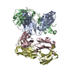
| ||||||||
|---|---|---|---|---|---|---|---|---|---|
| 1 | 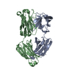
| ||||||||
| 2 | 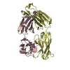
| ||||||||
| Unit cell |
| ||||||||
| Atom site foot note | 1: RESIDUE 141 IN CHAINS *A* AND *C* ARE CIS PROLINES. 2: RESIDUES 155 AND 157 IN CHAINS *B* AND *D* ARE CIS PROLINES. |
-Components
| #1: Antibody | Mass: 22767.197 Da / Num. of mol.: 2 / Source method: isolated from a natural source / Source: (natural) Homo sapiens (human) / References: PIR: S25738#2: Antibody | Mass: 24090.219 Da / Num. of mol.: 2 / Source method: isolated from a natural source / Source: (natural) Homo sapiens (human) / References: EMBL: Y14737#3: Water | ChemComp-HOH / |  Mass: 18.015 Da / Num. of mol.: 668 / Source method: isolated from a natural source / Formula: H2O Mass: 18.015 Da / Num. of mol.: 668 / Source method: isolated from a natural source / Formula: H2OHas protein modification | Y | Sequence details | IN THE SEQUENCE DATA BASE, THE SEQUENCE FOR THE HEAVY CHAIN IS INCOMPLETE AND THE SEQUENCE FOR THE ...IN THE SEQUENCE DATA BASE, THE SEQUENCE FOR THE HEAVY CHAIN IS INCOMPLETE | |
|---|
-Experimental details
-Experiment
| Experiment | Method: X-RAY DIFFRACTION |
|---|
- Sample preparation
| Crystal | Density Matthews: 2.5 Å3/Da / Density % sol: 50.77 % | ||||||||||||||||||||||||||||||||||||||||||||||||
|---|---|---|---|---|---|---|---|---|---|---|---|---|---|---|---|---|---|---|---|---|---|---|---|---|---|---|---|---|---|---|---|---|---|---|---|---|---|---|---|---|---|---|---|---|---|---|---|---|---|
| Crystal grow | *PLUS Method: vapor diffusionDetails: referred to 'Rose,D.R.', (1990) Proc.Natl.Acad.Sci.U.S.A., 87, 338-342 | ||||||||||||||||||||||||||||||||||||||||||||||||
| Components of the solutions | *PLUS
|
- Processing
| Software |
| ||||||||||||||||||||||||||||||||||||||||||||||||||||||||||||
|---|---|---|---|---|---|---|---|---|---|---|---|---|---|---|---|---|---|---|---|---|---|---|---|---|---|---|---|---|---|---|---|---|---|---|---|---|---|---|---|---|---|---|---|---|---|---|---|---|---|---|---|---|---|---|---|---|---|---|---|---|---|
| Refinement | Resolution: 1.8→6 Å / σ(F): 2.5 /
| ||||||||||||||||||||||||||||||||||||||||||||||||||||||||||||
| Refinement step | Cycle: LAST / Resolution: 1.8→6 Å
| ||||||||||||||||||||||||||||||||||||||||||||||||||||||||||||
| Refine LS restraints |
| ||||||||||||||||||||||||||||||||||||||||||||||||||||||||||||
| Refinement | *PLUS Highest resolution: 1.8 Å / Lowest resolution: 6 Å / Num. reflection obs: 64477 / σ(F): 2.5 / Rfactor obs: 0.173 | ||||||||||||||||||||||||||||||||||||||||||||||||||||||||||||
| Solvent computation | *PLUS | ||||||||||||||||||||||||||||||||||||||||||||||||||||||||||||
| Displacement parameters | *PLUS | ||||||||||||||||||||||||||||||||||||||||||||||||||||||||||||
| Refine LS restraints | *PLUS Type: x_angle_d / Dev ideal: 2.96 |
