 Movie
Movie Controller

[English] 日本語
 Yorodumi
- PDB-6tul: Structure of the arginase-2-inhibitory human antigen-binding frag... -
+ Open data

- Basic information
| Entry | Database: PDB / ID: 6tul | ||||||
|---|---|---|---|---|---|---|---|
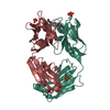
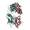
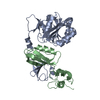
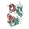
| Title | Structure of the arginase-2-inhibitory human antigen-binding fragment Fab C0021177 | ||||||
 Components Components | (Fab C0021177 ...) x 2 | ||||||
 Keywords Keywords | PROTEIN BINDING / arginase-2 inhibitor / IgG / antigen-binding fragment | ||||||
| Function / homology | D-MALATE / TRIETHYLENE GLYCOL Function and homology information Function and homology information | ||||||
| Biological species |  Homo sapiens (human) Homo sapiens (human) | ||||||
| Method |  X-RAY DIFFRACTION / SYNCHROTRON / MOLECULAR REPLACEMENT / Resolution: 2.25 Å X-RAY DIFFRACTION / SYNCHROTRON / MOLECULAR REPLACEMENT / Resolution: 2.25 Å | ||||||
 Authors Authors | Burschowsky, D. / Addyman, A. / Fiedler, S. / Groves, M. / Haynes, S. / Seewooruthun, C. / Carr, M. | ||||||
| Funding support |  United Kingdom, 1items United Kingdom, 1items
| ||||||
 Citation Citation | Journal: Mabs Title: Structural and functional characterization of C0021158, a high-affinity monoclonal antibody that inhibits Arginase 2 function via a novel non-competitive mechanism of action. Authors: Austin, M. / Burschowsky, D. / Chan, D.T.Y. / Jenkinson, L. / Haynes, S. / Diamandakis, A. / Seewooruthun, C. / Addyman, A. / Fiedler, S. / Ryman, S. / Whitehouse, J. / Slater, L.H. / ...Authors: Austin, M. / Burschowsky, D. / Chan, D.T.Y. / Jenkinson, L. / Haynes, S. / Diamandakis, A. / Seewooruthun, C. / Addyman, A. / Fiedler, S. / Ryman, S. / Whitehouse, J. / Slater, L.H. / Hadjinicolaou, A.V. / Gileadi, U. / Gowans, E. / Shibata, Y. / Barnard, M. / Kaserer, T. / Sharma, P. / Luheshi, N.M. / Wilkinson, R.W. / Vaughan, T.J. / Holt, S.V. / Cerundolo, V. / Carr, M.D. / Groves, M.A.T. | ||||||
| History |
|
- Structure visualization
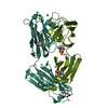
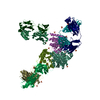
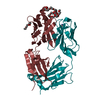
| Structure viewer | Molecule: MolmilJmol/JSmol |
|---|
- Downloads & links
-Download
| PDBx/mmCIF format | 6tul.cif.gz | 303 KB | Display | PDBx/mmCIF format |
|---|---|---|---|---|
| PDB format | pdb6tul.ent.gz | Display | PDB format | |
| PDBx/mmJSON format | 6tul.json.gz | Tree view | PDBx/mmJSON format | |
| Others |  Other downloads Other downloads |
-Validation report
| Arichive directory | https://data.pdbj.org/pub/pdb/validation_reports/tu/6tulftp://data.pdbj.org/pub/pdb/validation_reports/tu/6tul | HTTPS FTP |
|---|
-Related structure data
| Related structure data | 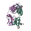 6srvSC 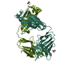 6srxC 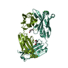 6ss0C 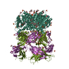 6ss2C 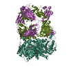 6ss4C S: Starting model for refinement C: citing same article ( |
|---|---|
| Similar structure data |


-Links
 PDBj
PDBj


- Assembly
| Deposited unit | 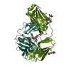
| ||||||||
|---|---|---|---|---|---|---|---|---|---|
| 1 |
| ||||||||
| Unit cell |
|
-Components
-Antibody , 2 types, 2 molecules HHHLLL
| #1: Antibody | Mass: 24605.717 Da / Num. of mol.: 1 Source method: isolated from a genetically manipulated source Details: Amino acids are numbered according to the Kabat numbering scheme. Source: (gene. exp.) Homo sapiens (human) / Plasmid: pEU1.3 fab / Cell line (production host): CHO / Production host:   Cricetulus griseus (Chinese hamster) Cricetulus griseus (Chinese hamster) |
|---|---|
| #2: Antibody | Mass: 23096.484 Da / Num. of mol.: 1 Source method: isolated from a genetically manipulated source Details: Amino acids are numbered according to the Kabat numbering scheme. Residue 10 would not be present in the Kabat scheme, but due to problems with non-continuous numbering in L-peptides, we ...Details: Amino acids are numbered according to the Kabat numbering scheme. Residue 10 would not be present in the Kabat scheme, but due to problems with non-continuous numbering in L-peptides, we included it. Therefore, residues 2-10 should be read as 1-9, per Kabat. Source: (gene. exp.) Homo sapiens (human) / Plasmid: pEU1.3 fab / Cell line (production host): CHO / Production host: Cricetulus griseus (Chinese hamster) |
-Non-polymers , 5 types, 52 molecules 


| #3: Chemical |  Mass: 194.226 Da / Num. of mol.: 2 / Source method: obtained synthetically / Formula: C8H18O5 / Comment: precipitant*YM Mass: 194.226 Da / Num. of mol.: 2 / Source method: obtained synthetically / Formula: C8H18O5 / Comment: precipitant*YM#4: Chemical | ChemComp-P6G / |  Mass: 282.331 Da / Num. of mol.: 1 / Source method: obtained synthetically / Formula: C12H26O7 / Comment: precipitant*YM Mass: 282.331 Da / Num. of mol.: 1 / Source method: obtained synthetically / Formula: C12H26O7 / Comment: precipitant*YM#5: Chemical |  Mass: 134.087 Da / Num. of mol.: 2 / Source method: obtained synthetically / Formula: C4H6O5 Mass: 134.087 Da / Num. of mol.: 2 / Source method: obtained synthetically / Formula: C4H6O5#6: Chemical |  Mass: 150.173 Da / Num. of mol.: 2 / Source method: obtained synthetically / Formula: C6H14O4 Mass: 150.173 Da / Num. of mol.: 2 / Source method: obtained synthetically / Formula: C6H14O4#7: Water | ChemComp-HOH / | Mass: 18.015 Da / Num. of mol.: 45 / Source method: isolated from a natural source / Formula: H2O |
|---|
-Details
| Has ligand of interest | N |
|---|---|
| Has protein modification | Y |
-Experimental details
-Experiment
| Experiment | Method: X-RAY DIFFRACTION / Number of used crystals: 1 |
|---|
- Sample preparation
| Crystal | Density Matthews: 2.17 Å3/Da / Density % sol: 43.29 % |
|---|---|
| Crystal grow | Temperature: 291 K / Method: vapor diffusion, sitting drop / pH: 7 / Details: 100 mM MMT (D/L-malic acid, MES, tris) 25% PEG1500 |
-Data collection
| Diffraction | Mean temperature: 100 K / Serial crystal experiment: N |
|---|---|
| Diffraction source | Source: SYNCHROTRON / Site: BESSY  / Beamline: 14.1 / Wavelength: 0.9184 Å / Beamline: 14.1 / Wavelength: 0.9184 Å |
| Detector | Type: DECTRIS PILATUS 6M-F / Detector: PIXEL / Date: Sep 25, 2019 |
| Radiation | Protocol: SINGLE WAVELENGTH / Monochromatic (M) / Laue (L): M / Scattering type: x-ray |
| Radiation wavelength | Wavelength: 0.9184 Å / Relative weight: 1 |
| Reflection | Resolution: 2.25→47.83 Å / Num. obs: 19456 / % possible obs: 100 % / Redundancy: 12.8 % / CC1/2: 1 / Rmerge(I) obs: 0.18 / Net I/σ(I): 8.4 |
| Reflection shell | Resolution: 2.25→2.32 Å / Redundancy: 13.3 % / Rmerge(I) obs: 4.04 / Mean I/σ(I) obs: 0.7 / Num. unique obs: 1755 / CC1/2: 0.36 / % possible all: 99.9 |
- Processing
| Software |
| |||||||||||||||||||||||||||||||||||||||||||||||||||||||||||||||||||||||||||||||||||||||||||||||||||||||||||||||||||||||||||||||||||||||||||||||||||||||||||||||||||||||||||||||||||||||||||||||||||||||||||||||||||||||||||||||||||||||
|---|---|---|---|---|---|---|---|---|---|---|---|---|---|---|---|---|---|---|---|---|---|---|---|---|---|---|---|---|---|---|---|---|---|---|---|---|---|---|---|---|---|---|---|---|---|---|---|---|---|---|---|---|---|---|---|---|---|---|---|---|---|---|---|---|---|---|---|---|---|---|---|---|---|---|---|---|---|---|---|---|---|---|---|---|---|---|---|---|---|---|---|---|---|---|---|---|---|---|---|---|---|---|---|---|---|---|---|---|---|---|---|---|---|---|---|---|---|---|---|---|---|---|---|---|---|---|---|---|---|---|---|---|---|---|---|---|---|---|---|---|---|---|---|---|---|---|---|---|---|---|---|---|---|---|---|---|---|---|---|---|---|---|---|---|---|---|---|---|---|---|---|---|---|---|---|---|---|---|---|---|---|---|---|---|---|---|---|---|---|---|---|---|---|---|---|---|---|---|---|---|---|---|---|---|---|---|---|---|---|---|---|---|---|---|---|---|---|---|---|---|---|---|---|---|---|---|---|---|---|---|---|---|
| Refinement | Method to determine structure: MOLECULAR REPLACEMENT Starting model: 6SRV Resolution: 2.25→47.505 Å / Cor.coef. Fo:Fc: 0.944 / Cor.coef. Fo:Fc free: 0.899 / SU B: 36.359 / SU ML: 0.395 / Cross valid method: FREE R-VALUE / ESU R: 0.459 / ESU R Free: 0.309 Details: Hydrogens have been added in their riding positions
| |||||||||||||||||||||||||||||||||||||||||||||||||||||||||||||||||||||||||||||||||||||||||||||||||||||||||||||||||||||||||||||||||||||||||||||||||||||||||||||||||||||||||||||||||||||||||||||||||||||||||||||||||||||||||||||||||||||||
| Solvent computation | Ion probe radii: 0.8 Å / Shrinkage radii: 0.8 Å / VDW probe radii: 1.2 Å | |||||||||||||||||||||||||||||||||||||||||||||||||||||||||||||||||||||||||||||||||||||||||||||||||||||||||||||||||||||||||||||||||||||||||||||||||||||||||||||||||||||||||||||||||||||||||||||||||||||||||||||||||||||||||||||||||||||||
| Displacement parameters | Biso mean: 62.164 Å2
| |||||||||||||||||||||||||||||||||||||||||||||||||||||||||||||||||||||||||||||||||||||||||||||||||||||||||||||||||||||||||||||||||||||||||||||||||||||||||||||||||||||||||||||||||||||||||||||||||||||||||||||||||||||||||||||||||||||||
| Refinement step | Cycle: LAST / Resolution: 2.25→47.505 Å
| |||||||||||||||||||||||||||||||||||||||||||||||||||||||||||||||||||||||||||||||||||||||||||||||||||||||||||||||||||||||||||||||||||||||||||||||||||||||||||||||||||||||||||||||||||||||||||||||||||||||||||||||||||||||||||||||||||||||
| Refine LS restraints |
| |||||||||||||||||||||||||||||||||||||||||||||||||||||||||||||||||||||||||||||||||||||||||||||||||||||||||||||||||||||||||||||||||||||||||||||||||||||||||||||||||||||||||||||||||||||||||||||||||||||||||||||||||||||||||||||||||||||||
| LS refinement shell | Refine-ID: X-RAY DIFFRACTION / Total num. of bins used: 20
| |||||||||||||||||||||||||||||||||||||||||||||||||||||||||||||||||||||||||||||||||||||||||||||||||||||||||||||||||||||||||||||||||||||||||||||||||||||||||||||||||||||||||||||||||||||||||||||||||||||||||||||||||||||||||||||||||||||||
| Refinement TLS params. | Method: refined / Refine-ID: X-RAY DIFFRACTION
| |||||||||||||||||||||||||||||||||||||||||||||||||||||||||||||||||||||||||||||||||||||||||||||||||||||||||||||||||||||||||||||||||||||||||||||||||||||||||||||||||||||||||||||||||||||||||||||||||||||||||||||||||||||||||||||||||||||||
| Refinement TLS group |
|
