 Movie
Movie Controller

[English] 日本語
 Yorodumi
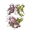
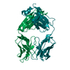
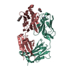
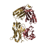
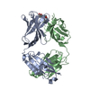
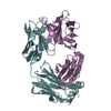
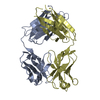
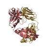
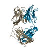
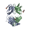
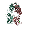
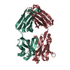
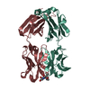
- PDB-4a6y: CRYSTAL STRUCTURE OF FAB FRAGMENT OF ANTI-(4-HYDROXY-3-NITROPHENY... -
+ Open data

- Basic information
| Entry | Database: PDB / ID: 4a6y | ||||||
|---|---|---|---|---|---|---|---|
| Title | CRYSTAL STRUCTURE OF FAB FRAGMENT OF ANTI-(4-HYDROXY-3-NITROPHENYL) -ACETYL MURINE GERMLINE ANTIBODY BBE6.12H3 | ||||||
 Components Components |
| ||||||
 Keywords Keywords | IMMUNE SYSTEM / ANTIBODY MULTISPECIFICITY / HUMORAL IMMUNE SYSTEM / COMPLEMENTARITY DETERMINING REGION FLEXIBILITY | ||||||
| Function / homology | Immunoglobulins / Immunoglobulin-like / Sandwich / Mainly Beta Function and homology information Function and homology information | ||||||
| Biological species |  | ||||||
| Method |  X-RAY DIFFRACTION / MOLECULAR REPLACEMENT / Resolution: 2.9 Å X-RAY DIFFRACTION / MOLECULAR REPLACEMENT / Resolution: 2.9 Å | ||||||
 Authors Authors | Khan, T. / Salunke, D.M. | ||||||
 Citation Citation | Journal: J.Immunol. / Year: 2012 Title: Structural Elucidation of the Mechanistic Basis of Degeneracy in the Primary Humoral Response. Authors: Khan, T. / Salunke, D.M. | ||||||
| History |
|
- Structure visualization
| Structure viewer | Molecule: MolmilJmol/JSmol |
|---|
- Downloads & links
-Download
| PDBx/mmCIF format | 4a6y.cif.gz | 172.6 KB | Display | PDBx/mmCIF format |
|---|---|---|---|---|
| PDB format | pdb4a6y.ent.gz | 137.3 KB | Display | PDB format |
| PDBx/mmJSON format | 4a6y.json.gz | Tree view | PDBx/mmJSON format | |
| Others |  Other downloads Other downloads |
-Validation report
| Arichive directory | https://data.pdbj.org/pub/pdb/validation_reports/a6/4a6yftp://data.pdbj.org/pub/pdb/validation_reports/a6/4a6y | HTTPS FTP |
|---|
-Related structure data
| Related structure data |  2xzqC  2y06C  2y07C  2y36C  1ngqS S: Starting model for refinement C: citing same article ( |
|---|---|
| Similar structure data |


-Links
 PDBj
PDBj

- Assembly
| Deposited unit | 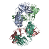
| ||||||||
|---|---|---|---|---|---|---|---|---|---|
| 1 | 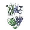
| ||||||||
| 2 | 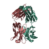
| ||||||||
| Unit cell |
|
-Components
| #1: Antibody | Mass: 22681.086 Da / Num. of mol.: 1 / Fragment: ANTIGEN BINDING / Source method: isolated from a natural source / Source: (natural) |
|---|---|
| #2: Antibody | Mass: 23748.617 Da / Num. of mol.: 1 / Fragment: ANTIGEN BINDING / Source method: isolated from a natural source / Source: (natural) |
| #3: Antibody | Mass: 23762.643 Da / Num. of mol.: 1 / Fragment: ANTIGEN BINDING / Source method: isolated from a natural source / Source: (natural) |
| #4: Antibody | Mass: 22695.174 Da / Num. of mol.: 1 / Fragment: ANTIGEN BINDING / Source method: isolated from a natural source / Source: (natural) |
| #5: Water | ChemComp-HOH /  Mass: 18.015 Da / Num. of mol.: 130 / Source method: isolated from a natural source / Formula: H2O Mass: 18.015 Da / Num. of mol.: 130 / Source method: isolated from a natural source / Formula: H2O |
| Has protein modification | Y |
-Experimental details
-Experiment
| Experiment | Method: X-RAY DIFFRACTION / Number of used crystals: 1 |
|---|
- Sample preparation
| Crystal | Density Matthews: 2.3 Å3/Da / Density % sol: 45.7 % / Description: NONE |
|---|---|
| Crystal grow | pH: 7.1 / Details: 21% PEG 3350 IN 0.05 M TRIS, PH 7.1 |
-Data collection
| Diffraction | Mean temperature: 120 K |
|---|---|
| Diffraction source | Source: ROTATING ANODE / Type: RIGAKU RU300 / Wavelength: 1.5418 |
| Detector | Type: MARRESEARCH / Detector: IMAGE PLATE / Date: Jun 15, 2006 / Details: MIRRORS |
| Radiation | Monochromator: NI FILTER CMF12 38CU-6 / Protocol: SINGLE WAVELENGTH / Monochromatic (M) / Laue (L): M / Scattering type: x-ray |
| Radiation wavelength | Wavelength: 1.5418 Å / Relative weight: 1 |
| Reflection | Resolution: 2.9→50 Å / Num. obs: 19688 / % possible obs: 93.5 % / Observed criterion σ(I): 0 / Redundancy: 3 % / Biso Wilson estimate: 45.7 Å2 / Rmerge(I) obs: 0.12 / Net I/σ(I): 4.7 |
| Reflection shell | Resolution: 2.9→3.08 Å / Redundancy: 2.6 % / Rmerge(I) obs: 0.42 / Mean I/σ(I) obs: 2.6 / % possible all: 87.3 |
- Processing
| Software |
| ||||||||||||||||||||||||||||||||||||||||||||||||||||||||||||||||||||||||||||||||
|---|---|---|---|---|---|---|---|---|---|---|---|---|---|---|---|---|---|---|---|---|---|---|---|---|---|---|---|---|---|---|---|---|---|---|---|---|---|---|---|---|---|---|---|---|---|---|---|---|---|---|---|---|---|---|---|---|---|---|---|---|---|---|---|---|---|---|---|---|---|---|---|---|---|---|---|---|---|---|---|---|---|
| Refinement | Method to determine structure: MOLECULAR REPLACEMENT Starting model: PDB ENTRY 1NGQ Resolution: 2.9→23.02 Å / Rfactor Rfree error: 0.008 / Data cutoff high absF: 125478.2 / Data cutoff low absF: 0 / Isotropic thermal model: RESTRAINED / Cross valid method: THROUGHOUT / σ(F): 0
| ||||||||||||||||||||||||||||||||||||||||||||||||||||||||||||||||||||||||||||||||
| Solvent computation | Solvent model: FLAT MODEL / Bsol: 29.3416 Å2 / ksol: 0.35 e/Å3 | ||||||||||||||||||||||||||||||||||||||||||||||||||||||||||||||||||||||||||||||||
| Displacement parameters | Biso mean: 31.2 Å2
| ||||||||||||||||||||||||||||||||||||||||||||||||||||||||||||||||||||||||||||||||
| Refine analyze |
| ||||||||||||||||||||||||||||||||||||||||||||||||||||||||||||||||||||||||||||||||
| Refinement step | Cycle: LAST / Resolution: 2.9→23.02 Å
| ||||||||||||||||||||||||||||||||||||||||||||||||||||||||||||||||||||||||||||||||
| Refine LS restraints |
| ||||||||||||||||||||||||||||||||||||||||||||||||||||||||||||||||||||||||||||||||
| Refine LS restraints NCS | NCS model details: NONE | ||||||||||||||||||||||||||||||||||||||||||||||||||||||||||||||||||||||||||||||||
| LS refinement shell | Resolution: 2.9→3.08 Å / Rfactor Rfree error: 0.024 / Total num. of bins used: 6
| ||||||||||||||||||||||||||||||||||||||||||||||||||||||||||||||||||||||||||||||||
| Xplor file |
|
