 Movie
Movie Controller

[English] 日本語
 Yorodumi
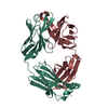
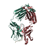
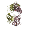
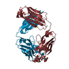
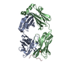
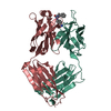
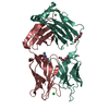
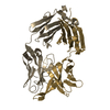
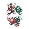
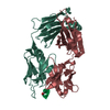
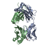
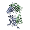
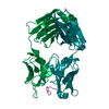
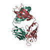
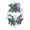
- PDB-2y06: CRYSTAL STRUCTURE ANALYSIS OF THE ANTI-(4-HYDROXY-3-NITROPHENYL) ... -
+ Open data

- Basic information
| Entry | Database: PDB / ID: 2y06 | ||||||
|---|---|---|---|---|---|---|---|
| Title | CRYSTAL STRUCTURE ANALYSIS OF THE ANTI-(4-HYDROXY-3-NITROPHENYL) - ACETYL MURINE GERMLINE ANTIBODY BBE6.12H3 FAB FRAGMENT IN COMPLEX WITH A PHAGE DISPLAY DERIVED DODECAPEPTIDE GDPRPSYISHLL | ||||||
 Components Components |
| ||||||
 Keywords Keywords | IMMUNE SYSTEM / BETA-SANDWICH IMMUNOGLOBULIN FOLD / ANTIGEN BINDING / SECRETED PROTEIN | ||||||
| Function / homology | Immunoglobulins / Immunoglobulin-like / Sandwich / Mainly Beta Function and homology information Function and homology information | ||||||
| Biological species |  SYNTHETIC CONSTRUCT (others) | ||||||
| Method |  X-RAY DIFFRACTION / SYNCHROTRON / MOLECULAR REPLACEMENT / Resolution: 2.5 Å X-RAY DIFFRACTION / SYNCHROTRON / MOLECULAR REPLACEMENT / Resolution: 2.5 Å | ||||||
 Authors Authors | Khan, T. / Salunke, D.M. | ||||||
 Citation Citation | Journal: J.Immunol. / Year: 2012 Title: Structural Elucidation of the Mechanistic Basis of Degeneracy in the Primary Humoral Response. Authors: Khan, T. / Salunke, D.M. | ||||||
| History |
|
- Structure visualization
| Structure viewer | Molecule: MolmilJmol/JSmol |
|---|
- Downloads & links
-Download
| PDBx/mmCIF format | 2y06.cif.gz | 95.2 KB | Display | PDBx/mmCIF format |
|---|---|---|---|---|
| PDB format | pdb2y06.ent.gz | 72.3 KB | Display | PDB format |
| PDBx/mmJSON format | 2y06.json.gz | Tree view | PDBx/mmJSON format | |
| Others |  Other downloads Other downloads |
-Validation report
| Arichive directory | https://data.pdbj.org/pub/pdb/validation_reports/y0/2y06ftp://data.pdbj.org/pub/pdb/validation_reports/y0/2y06 | HTTPS FTP |
|---|
-Related structure data


-Links
 PDBj
PDBj

- Assembly
| Deposited unit | 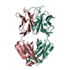
| ||||||||
|---|---|---|---|---|---|---|---|---|---|
| 1 |
| ||||||||
| Unit cell |
|
-Components
| #1: Antibody | Mass: 23458.199 Da / Num. of mol.: 1 / Source method: isolated from a natural source / Source: (natural) |
|---|---|
| #2: Antibody | Mass: 22538.885 Da / Num. of mol.: 1 / Source method: isolated from a natural source / Source: (natural) |
| #3: Protein/peptide | Mass: 1356.528 Da / Num. of mol.: 1 / Fragment: PEPTIDE, RESIDUES 1-12 / Source method: obtained synthetically / Source: (synth.) SYNTHETIC CONSTRUCT (others) |
| #4: Water | ChemComp-HOH /  Mass: 18.015 Da / Num. of mol.: 45 / Source method: isolated from a natural source / Formula: H2O Mass: 18.015 Da / Num. of mol.: 45 / Source method: isolated from a natural source / Formula: H2O |
| Has protein modification | Y |
-Experimental details
-Experiment
| Experiment | Method: X-RAY DIFFRACTION / Number of used crystals: 1 |
|---|
- Sample preparation
| Crystal | Density Matthews: 1.93 Å3/Da / Density % sol: 36.46 % / Description: NONE |
|---|---|
| Crystal grow | Temperature: 301 K / Method: vapor diffusion, hanging drop / pH: 7.1 Details: 0.05 M TRIS HCL, 0.025% SODIUM AZIDE, PEG 8000., pH 7.1 |
-Data collection
| Diffraction | Mean temperature: 288 K |
|---|---|
| Diffraction source | Source: SYNCHROTRON / Site: ESRF  / Beamline: BM14 / Wavelength: 0.97 / Beamline: BM14 / Wavelength: 0.97 |
| Detector | Type: MARMOSAIC 225 mm CCD / Detector: CCD / Date: Apr 12, 2009 / Details: BENT COLLIMATING MIRROR AND TOROID |
| Radiation | Monochromator: SI(111) MONOCHROMATOR / Protocol: SINGLE WAVELENGTH / Monochromatic (M) / Laue (L): M / Scattering type: x-ray |
| Radiation wavelength | Wavelength: 0.97 Å / Relative weight: 1 |
| Reflection | Resolution: 2.5→41.3 Å / Num. obs: 13312 / % possible obs: 99.8 % / Observed criterion σ(I): 0 / Redundancy: 3.9 % / Biso Wilson estimate: 59 Å2 / Rmerge(I) obs: 0.06 / Net I/σ(I): 10 |
| Reflection shell | Resolution: 2.5→7.9 Å / Redundancy: 3.7 % / Rmerge(I) obs: 0.23 / Mean I/σ(I) obs: 2.5 / % possible all: 99.3 |
- Processing
| Software |
| ||||||||||||||||||||||||||||||||||||||||||||||||||||||||||||
|---|---|---|---|---|---|---|---|---|---|---|---|---|---|---|---|---|---|---|---|---|---|---|---|---|---|---|---|---|---|---|---|---|---|---|---|---|---|---|---|---|---|---|---|---|---|---|---|---|---|---|---|---|---|---|---|---|---|---|---|---|---|
| Refinement | Method to determine structure: MOLECULAR REPLACEMENT Starting model: NATIVE FAB STRUCTURE Resolution: 2.5→41.3 Å / Rfactor Rfree error: 0.008 / Data cutoff high absF: 137292.07 / Data cutoff low absF: 0 / Cross valid method: THROUGHOUT / σ(F): 0 / Stereochemistry target values: MAXIMUM LIKELIHOOD
| ||||||||||||||||||||||||||||||||||||||||||||||||||||||||||||
| Solvent computation | Solvent model: FLAT MODEL / Bsol: 37.968 Å2 / ksol: 0.3 e/Å3 | ||||||||||||||||||||||||||||||||||||||||||||||||||||||||||||
| Displacement parameters | Biso mean: 68 Å2
| ||||||||||||||||||||||||||||||||||||||||||||||||||||||||||||
| Refine analyze | Luzzati coordinate error obs: 0.37 Å / Luzzati d res low obs: 5 Å / Luzzati sigma a obs: 0.34 Å | ||||||||||||||||||||||||||||||||||||||||||||||||||||||||||||
| Refinement step | Cycle: LAST / Resolution: 2.5→41.3 Å
| ||||||||||||||||||||||||||||||||||||||||||||||||||||||||||||
| Refine LS restraints |
| ||||||||||||||||||||||||||||||||||||||||||||||||||||||||||||
| LS refinement shell | Resolution: 2.5→2.66 Å / Rfactor Rfree error: 0.04 / Total num. of bins used: 24
| ||||||||||||||||||||||||||||||||||||||||||||||||||||||||||||
| Xplor file |
|
