 Movie
Movie Controller


+ Open data

- Basic information
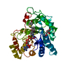
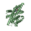
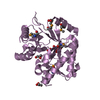
| Entry | Database: PDB / ID: 6unr | ||||||
|---|---|---|---|---|---|---|---|
| Title | Kinase domain of ALK2-K492A/K493A with AMPPNP | ||||||
 Components Components | Activin receptor type-1 | ||||||
 Keywords Keywords | TRANSFERASE / Kinase | ||||||
| Function / homology |  Function and homology information Function and homology informationendocardial cushion cell fate commitment / mitral valve morphogenesis / BMP receptor complex / BMP receptor activity / endocardial cushion fusion / cardiac muscle cell fate commitment / atrial septum primum morphogenesis / positive regulation of cardiac epithelial to mesenchymal transition / acute inflammatory response / positive regulation of determination of dorsal identity ...endocardial cushion cell fate commitment / mitral valve morphogenesis / BMP receptor complex / BMP receptor activity / endocardial cushion fusion / cardiac muscle cell fate commitment / atrial septum primum morphogenesis / positive regulation of cardiac epithelial to mesenchymal transition / acute inflammatory response / positive regulation of determination of dorsal identity / transforming growth factor beta receptor activity, type I / smooth muscle cell differentiation / activin receptor activity, type I / activin receptor complex / endocardial cushion formation / pharyngeal system development / receptor protein serine/threonine kinase / activin binding / cellular response to BMP stimulus / transmembrane receptor protein serine/threonine kinase activity / activin receptor signaling pathway / negative regulation of activin receptor signaling pathway / embryonic heart tube morphogenesis / dorsal/ventral pattern formation / gastrulation with mouth forming second / transforming growth factor beta binding / determination of left/right symmetry / atrioventricular valve morphogenesis / neural crest cell migration / branching involved in blood vessel morphogenesis / ventricular septum morphogenesis / negative regulation of G1/S transition of mitotic cell cycle / SMAD binding / germ cell development / peptide hormone binding / mesoderm formation / positive regulation of SMAD protein signal transduction / positive regulation of intracellular signal transduction / regulation of ossification / positive regulation of osteoblast differentiation / positive regulation of bone mineralization / negative regulation of signal transduction / BMP signaling pathway / transforming growth factor beta receptor signaling pathway / protein tyrosine kinase binding / negative regulation of extrinsic apoptotic signaling pathway / cellular response to growth factor stimulus / apical part of cell / osteoblast differentiation / heart development / in utero embryonic development / cell differentiation / protein kinase activity / positive regulation of cell migration / cadherin binding / protein serine/threonine kinase activity / positive regulation of DNA-templated transcription / protein homodimerization activity / positive regulation of transcription by RNA polymerase II / ATP binding / metal ion binding / plasma membrane Similarity search - Function | ||||||
| Biological species |  Homo sapiens (human) Homo sapiens (human) | ||||||
| Method |  X-RAY DIFFRACTION / SYNCHROTRON / MOLECULAR REPLACEMENT / Resolution: 2.2 Å X-RAY DIFFRACTION / SYNCHROTRON / MOLECULAR REPLACEMENT / Resolution: 2.2 Å | ||||||
 Authors Authors | Agnew, C. / Jura, N. | ||||||
 Citation Citation | Journal: Nat Commun / Year: 2021 Title: Structural basis for ALK2/BMPR2 receptor complex signaling through kinase domain oligomerization. Authors: Agnew, C. / Ayaz, P. / Kashima, R. / Loving, H.S. / Ghatpande, P. / Kung, J.E. / Underbakke, E.S. / Shan, Y. / Shaw, D.E. / Hata, A. / Jura, N. | ||||||
| History |
|
- Structure visualization
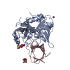
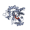
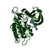
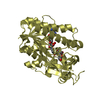
| Structure viewer | Molecule: MolmilJmol/JSmol |
|---|
- Downloads & links
-Download
| PDBx/mmCIF format | 6unr.cif.gz | 157.3 KB | Display | PDBx/mmCIF format |
|---|---|---|---|---|
| PDB format | pdb6unr.ent.gz | 101 KB | Display | PDB format |
| PDBx/mmJSON format | 6unr.json.gz | Tree view | PDBx/mmJSON format | |
| Others |  Other downloads Other downloads |
-Validation report
| Arichive directory | https://data.pdbj.org/pub/pdb/validation_reports/un/6unrftp://data.pdbj.org/pub/pdb/validation_reports/un/6unr | HTTPS FTP |
|---|
-Related structure data
| Related structure data | 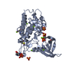 6unpC  6unqC 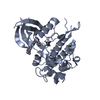 6unsC  3q4uS C: citing same article ( S: Starting model for refinement |
|---|---|
| Similar structure data |


-Links
 PDBj
PDBj


- Assembly
| Deposited unit | 
| ||||||||||||
|---|---|---|---|---|---|---|---|---|---|---|---|---|---|
| 1 |
| ||||||||||||
| Unit cell |
|
-Components
| #1: Protein | Mass: 38059.336 Da / Num. of mol.: 1 / Fragment: Kinase domain / Mutation: K492A, K493A Source method: isolated from a genetically manipulated source Source: (gene. exp.) Homo sapiens (human) / Gene: ACVR1, ACVRLK2 / Production host:   Spodoptera frugiperda (fall armyworm) Spodoptera frugiperda (fall armyworm)References: UniProt: Q04771, receptor protein serine/threonine kinase | ||||
|---|---|---|---|---|---|
| #2: Chemical | ChemComp-ANP /   Mass: 506.196 Da / Num. of mol.: 1 / Source method: obtained synthetically / Formula: C10H17N6O12P3 / Comment: AMP-PNP, energy-carrying molecule analogue*YM Mass: 506.196 Da / Num. of mol.: 1 / Source method: obtained synthetically / Formula: C10H17N6O12P3 / Comment: AMP-PNP, energy-carrying molecule analogue*YM | ||||
| #3: Chemical | ChemComp-MG /   Mass: 24.305 Da / Num. of mol.: 8 / Source method: obtained synthetically / Formula: Mg Mass: 24.305 Da / Num. of mol.: 8 / Source method: obtained synthetically / Formula: Mg#4: Water | ChemComp-HOH / |  Mass: 18.015 Da / Num. of mol.: 12 / Source method: isolated from a natural source / Formula: H2O Mass: 18.015 Da / Num. of mol.: 12 / Source method: isolated from a natural source / Formula: H2OHas ligand of interest | N | |
-Experimental details
-Experiment
| Experiment | Method: X-RAY DIFFRACTION / Number of used crystals: 1 |
|---|
- Sample preparation
| Crystal | Density Matthews: 2.35 Å3/Da / Density % sol: 47.66 % |
|---|---|
| Crystal grow | Temperature: 298 K / Method: vapor diffusion, sitting drop / pH: 7 / Details: 0.05 M PIPES 7 pH, 0.01 M DTT, 10 %w/v PEG 4000 |
-Data collection
| Diffraction | Mean temperature: 80 K / Serial crystal experiment: N |
|---|---|
| Diffraction source | Source: SYNCHROTRON / Site: ALS  / Beamline: 8.3.1 / Wavelength: 1.1111 Å / Beamline: 8.3.1 / Wavelength: 1.1111 Å |
| Detector | Type: DECTRIS PILATUS 6M / Detector: PIXEL / Date: Jun 27, 2018 |
| Radiation | Protocol: SINGLE WAVELENGTH / Monochromatic (M) / Laue (L): M / Scattering type: x-ray |
| Radiation wavelength | Wavelength: 1.1111 Å / Relative weight: 1 |
| Reflection | Resolution: 2.2→48.92 Å / Num. obs: 18605 / % possible obs: 99.9 % / Redundancy: 7.2 % / Biso Wilson estimate: 42.4 Å2 / CC1/2: 0.99 / Rmerge(I) obs: 0.102 / Rpim(I) all: 0.06 / Rrim(I) all: 0.119 / Net I/σ(I): 10.6 |
| Reflection shell | Resolution: 2.2→2.27 Å / Rmerge(I) obs: 1.172 / Mean I/σ(I) obs: 1.9 / Num. unique obs: 1571 / CC1/2: 0.929 / Rpim(I) all: 0.685 / Rrim(I) all: 1.361 |
- Processing
| Software |
| |||||||||||||||||||||||||||||||||||||||||||||||||
|---|---|---|---|---|---|---|---|---|---|---|---|---|---|---|---|---|---|---|---|---|---|---|---|---|---|---|---|---|---|---|---|---|---|---|---|---|---|---|---|---|---|---|---|---|---|---|---|---|---|---|
| Refinement | Method to determine structure: MOLECULAR REPLACEMENT Starting model: 3Q4U Resolution: 2.2→46.14 Å / SU ML: 0.3484 / Cross valid method: FREE R-VALUE / σ(F): 1.34 / Phase error: 39.7216 Stereochemistry target values: GeoStd + Monomer Library + CDL v1.2
| |||||||||||||||||||||||||||||||||||||||||||||||||
| Solvent computation | Shrinkage radii: 0.9 Å / VDW probe radii: 1.11 Å / Solvent model: FLAT BULK SOLVENT MODEL | |||||||||||||||||||||||||||||||||||||||||||||||||
| Displacement parameters | Biso mean: 74.05 Å2 | |||||||||||||||||||||||||||||||||||||||||||||||||
| Refinement step | Cycle: LAST / Resolution: 2.2→46.14 Å
| |||||||||||||||||||||||||||||||||||||||||||||||||
| Refine LS restraints |
| |||||||||||||||||||||||||||||||||||||||||||||||||
| LS refinement shell |
| |||||||||||||||||||||||||||||||||||||||||||||||||
| Refinement TLS params. | Method: refined / Origin x: 13.5349426149 Å / Origin y: 18.5918193248 Å / Origin z: -17.416687112 Å
| |||||||||||||||||||||||||||||||||||||||||||||||||
| Refinement TLS group | Selection details: all |
