 Movie
Movie Controller

[English] 日本語
 Yorodumi
- PDB-2c29: Structure of dihydroflavonol reductase from Vitis vinifera at 1.8 A. -
+ Open data

- Basic information
| Entry | Database: PDB / ID: 2c29 | ||||||
|---|---|---|---|---|---|---|---|
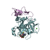
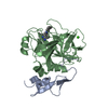
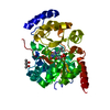
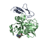
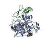
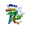
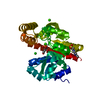
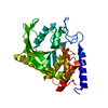
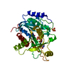
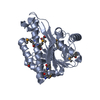
| Title | Structure of dihydroflavonol reductase from Vitis vinifera at 1.8 A. | ||||||
 Components Components | DIHYDROFLAVONOL 4-REDUCTASE | ||||||
 Keywords Keywords | OXIDOREDUCTASE / FLAVONOIDS / SHORT DEHYDROGENASE REDUCTASE / NADPH / DIHYDROQUERCETIN / ROSSMANN FOLD | ||||||
| Function / homology |  Function and homology information Function and homology informationdihydroflavonol 4-reductase / dihydroflavanol 4-reductase activity / flavanone 4-reductase / flavanone 4-reductase activity / flavonoid biosynthetic process / nucleotide binding Similarity search - Function | ||||||
| Biological species |  | ||||||
| Method |  X-RAY DIFFRACTION / SYNCHROTRON / MAD / Resolution: 1.81 Å X-RAY DIFFRACTION / SYNCHROTRON / MAD / Resolution: 1.81 Å | ||||||
 Authors Authors | Petit, P. / Granier, T. / D'Estaintot, B.L. / Hamdi, S. / Gallois, B. | ||||||
 Citation Citation | Journal: J.Mol.Biol. / Year: 2007 Title: Crystal Structure of Grape Dihydroflavonol 4-Reductase, a Key Enzyme in Flavonoid Biosynthesis. Authors: Petit, P. / Granier, T. / D'Estaintot, B.L. / Manigand, C. / Bathany, K. / Schmitter, J.M. / Lauvergeat, V. / Hamdi, S. / Gallois, B. | ||||||
| History |
|
- Structure visualization
| Structure viewer | Molecule: MolmilJmol/JSmol |
|---|
- Downloads & links
-Download
| PDBx/mmCIF format | 2c29.cif.gz | 152.1 KB | Display | PDBx/mmCIF format |
|---|---|---|---|---|
| PDB format | pdb2c29.ent.gz | 120.5 KB | Display | PDB format |
| PDBx/mmJSON format | 2c29.json.gz | Tree view | PDBx/mmJSON format | |
| Others |  Other downloads Other downloads |
-Validation report
| Arichive directory | https://data.pdbj.org/pub/pdb/validation_reports/c2/2c29ftp://data.pdbj.org/pub/pdb/validation_reports/c2/2c29 | HTTPS FTP |
|---|
-Related structure data
| Similar structure data |
|---|
-Links
 PDBj
PDBj- Assembly
| Deposited unit | 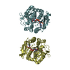
| |||||||||||||||||||||||||||||||||||||||||||||||||||||||||||||||||||||||||||
|---|---|---|---|---|---|---|---|---|---|---|---|---|---|---|---|---|---|---|---|---|---|---|---|---|---|---|---|---|---|---|---|---|---|---|---|---|---|---|---|---|---|---|---|---|---|---|---|---|---|---|---|---|---|---|---|---|---|---|---|---|---|---|---|---|---|---|---|---|---|---|---|---|---|---|---|---|
| 1 | 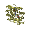
| |||||||||||||||||||||||||||||||||||||||||||||||||||||||||||||||||||||||||||
| 2 | 
| |||||||||||||||||||||||||||||||||||||||||||||||||||||||||||||||||||||||||||
| Unit cell |
| |||||||||||||||||||||||||||||||||||||||||||||||||||||||||||||||||||||||||||
| Noncrystallographic symmetry (NCS) | NCS domain:
NCS domain segments:
NCS oper: (Code: given Matrix: (-0.03694, -0.99926, 0.01094), Vector: |
-Components
| #1: Protein | Mass: 37694.344 Da / Num. of mol.: 2 Source method: isolated from a genetically manipulated source Details: ROSSMANN FOLD / Source: (gene. exp.)  #2: Chemical |   Mass: 743.405 Da / Num. of mol.: 2 / Source method: obtained synthetically / Formula: C21H28N7O17P3 Mass: 743.405 Da / Num. of mol.: 2 / Source method: obtained synthetically / Formula: C21H28N7O17P3#3: Chemical |   Mass: 304.252 Da / Num. of mol.: 2 / Source method: obtained synthetically / Formula: C15H12O7 Mass: 304.252 Da / Num. of mol.: 2 / Source method: obtained synthetically / Formula: C15H12O7#4: Water | ChemComp-HOH / |  Mass: 18.015 Da / Num. of mol.: 576 / Source method: isolated from a natural source / Formula: H2O Mass: 18.015 Da / Num. of mol.: 576 / Source method: isolated from a natural source / Formula: H2O |
|---|
-Experimental details
-Experiment
| Experiment | Method: X-RAY DIFFRACTION / Number of used crystals: 1 |
|---|
- Sample preparation
| Crystal | Density Matthews: 2.29 Å3/Da / Density % sol: 45.83 % |
|---|---|
| Crystal grow | pH: 7.5 / Details: NACL 80-200MM PEG 3350 25-30% HEPES 100MM PH 7.5 |
-Data collection
| Diffraction | Mean temperature: 100 K |
|---|---|
| Diffraction source | Source: SYNCHROTRON / Site: ESRF  / Beamline: BM30A / Wavelength: 0.97319 / Beamline: BM30A / Wavelength: 0.97319 |
| Detector | Type: MARRESEARCH / Detector: CCD / Date: Mar 5, 2005 / Details: MIRRORS |
| Radiation | Monochromator: SI (111) / Protocol: SINGLE WAVELENGTH / Monochromatic (M) / Laue (L): M / Scattering type: x-ray |
| Radiation wavelength | Wavelength: 0.97319 Å / Relative weight: 1 |
| Reflection | Resolution: 1.81→1.91 Å / Num. obs: 66981 / % possible obs: 98.9 % / Observed criterion σ(I): 2 / Redundancy: 5.3 % / Rmerge(I) obs: 0.07 / Net I/σ(I): 6.7 |
| Reflection shell | Resolution: 1.81→1.91 Å / Redundancy: 3.3 % / Rmerge(I) obs: 0.59 / Mean I/σ(I) obs: 2 / % possible all: 98.1 |
- Processing
| Software |
| ||||||||||||||||||||||||||||||||||||||||||||||||||||||||||||||||||||||||||||||||||||||||||||||||||||||||||||||||||||||||||||||||||||||||||||||||||||||||||||||||||||||||||||||||||||||
|---|---|---|---|---|---|---|---|---|---|---|---|---|---|---|---|---|---|---|---|---|---|---|---|---|---|---|---|---|---|---|---|---|---|---|---|---|---|---|---|---|---|---|---|---|---|---|---|---|---|---|---|---|---|---|---|---|---|---|---|---|---|---|---|---|---|---|---|---|---|---|---|---|---|---|---|---|---|---|---|---|---|---|---|---|---|---|---|---|---|---|---|---|---|---|---|---|---|---|---|---|---|---|---|---|---|---|---|---|---|---|---|---|---|---|---|---|---|---|---|---|---|---|---|---|---|---|---|---|---|---|---|---|---|---|---|---|---|---|---|---|---|---|---|---|---|---|---|---|---|---|---|---|---|---|---|---|---|---|---|---|---|---|---|---|---|---|---|---|---|---|---|---|---|---|---|---|---|---|---|---|---|---|---|
| Refinement | Method to determine structure: MAD / Resolution: 1.81→16.43 Å / Cor.coef. Fo:Fc: 0.964 / Cor.coef. Fo:Fc free: 0.939 / Cross valid method: THROUGHOUT / ESU R: 0.134 / ESU R Free: 0.136 / Stereochemistry target values: MAXIMUM LIKELIHOOD / Details: HYDROGENS HAVE BEEN ADDED IN THE RIDING POSITIONS.
| ||||||||||||||||||||||||||||||||||||||||||||||||||||||||||||||||||||||||||||||||||||||||||||||||||||||||||||||||||||||||||||||||||||||||||||||||||||||||||||||||||||||||||||||||||||||
| Solvent computation | Ion probe radii: 0.8 Å / Shrinkage radii: 0.8 Å / VDW probe radii: 1.2 Å / Solvent model: MASK | ||||||||||||||||||||||||||||||||||||||||||||||||||||||||||||||||||||||||||||||||||||||||||||||||||||||||||||||||||||||||||||||||||||||||||||||||||||||||||||||||||||||||||||||||||||||
| Displacement parameters | Biso mean: 28.43 Å2
| ||||||||||||||||||||||||||||||||||||||||||||||||||||||||||||||||||||||||||||||||||||||||||||||||||||||||||||||||||||||||||||||||||||||||||||||||||||||||||||||||||||||||||||||||||||||
| Refinement step | Cycle: LAST / Resolution: 1.81→16.43 Å
| ||||||||||||||||||||||||||||||||||||||||||||||||||||||||||||||||||||||||||||||||||||||||||||||||||||||||||||||||||||||||||||||||||||||||||||||||||||||||||||||||||||||||||||||||||||||
| Refine LS restraints |
|