 Movie
Movie Controller

[English] 日本語
 Yorodumi
- PDB-6dt6: Crystal Structure of the YopH PTP1B Chimera 3 PTPase bound to vanadate -
+ Open data

- Basic information
| Entry | Database: PDB / ID: 6dt6 | |||||||||
|---|---|---|---|---|---|---|---|---|---|---|
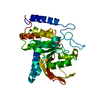
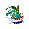
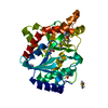
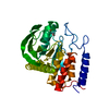
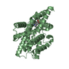
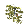
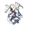
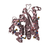
| Title | Crystal Structure of the YopH PTP1B Chimera 3 PTPase bound to vanadate | |||||||||
 Components Components | Targeted effector protein | |||||||||
 Keywords Keywords | HYDROLASE / PTPase / phosphatase / PTP / YopH | |||||||||
| Function / homology |  Function and homology information Function and homology informationsymbiont-mediated suppression of host reactive oxygen species generation / symbiont-mediated disruption of host focal adhesion / protein-tyrosine-phosphatase / protein tyrosine phosphatase activity / extracellular region Similarity search - Function | |||||||||
| Biological species |   Yersinia pestis (bacteria) Yersinia pestis (bacteria) | |||||||||
| Method |  X-RAY DIFFRACTION / SYNCHROTRON / MOLECULAR REPLACEMENT / Resolution: 2.101 Å X-RAY DIFFRACTION / SYNCHROTRON / MOLECULAR REPLACEMENT / Resolution: 2.101 Å | |||||||||
 Authors Authors | Morales, Y. / Johnson, S.J. / Hengge, A.C. | |||||||||
| Funding support |  United States, 2items United States, 2items
| |||||||||
 Citation Citation | Journal: Biochemistry / Year: 2018 Title: A YopH PTP1B Chimera Shows the Importance of the WPD-Loop Sequence to the Activity, Structure, and Dynamics of Protein Tyrosine Phosphatases. Authors: Moise, G. / Morales, Y. / Beaumont, V. / Caradonna, T. / Loria, J.P. / Johnson, S.J. / Hengge, A.C. | |||||||||
| History |
|
- Structure visualization
| Structure viewer | Molecule: MolmilJmol/JSmol |
|---|
- Downloads & links
-Download
| PDBx/mmCIF format | 6dt6.cif.gz | 126.6 KB | Display | PDBx/mmCIF format |
|---|---|---|---|---|
| PDB format | pdb6dt6.ent.gz | 96.1 KB | Display | PDB format |
| PDBx/mmJSON format | 6dt6.json.gz | Tree view | PDBx/mmJSON format | |
| Others |  Other downloads Other downloads |
-Validation report
| Arichive directory | https://data.pdbj.org/pub/pdb/validation_reports/dt/6dt6ftp://data.pdbj.org/pub/pdb/validation_reports/dt/6dt6 | HTTPS FTP |
|---|
-Related structure data
| Related structure data |  6dr1C  6dr7C  6dr9C  6drbC  4yaaS S: Starting model for refinement C: citing same article ( |
|---|---|
| Similar structure data |


-Links
 PDBj
PDBj


- Assembly
| Deposited unit | 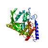
| ||||||||
|---|---|---|---|---|---|---|---|---|---|
| 1 |
| ||||||||
| Unit cell |
|
-Components
| #1: Protein | Mass: 33587.984 Da / Num. of mol.: 1 Source method: isolated from a genetically manipulated source Details: Residues 163-186 not visible / Source: (gene. exp.) Yersinia pestis (bacteria) / Gene: yopH, YPCD1.67c / Plasmid: pD-Nus1 / Production host: |
|---|---|
| #2: Chemical | ChemComp-VO4 /   Mass: 114.939 Da / Num. of mol.: 1 / Source method: obtained synthetically / Formula: VO4 Mass: 114.939 Da / Num. of mol.: 1 / Source method: obtained synthetically / Formula: VO4 |
| #3: Water | ChemComp-HOH /  Mass: 18.015 Da / Num. of mol.: 105 / Source method: isolated from a natural source / Formula: H2O Mass: 18.015 Da / Num. of mol.: 105 / Source method: isolated from a natural source / Formula: H2O |
-Experimental details
-Experiment
| Experiment | Method: X-RAY DIFFRACTION / Number of used crystals: 1 |
|---|
- Sample preparation
| Crystal | Density Matthews: 1.94 Å3/Da / Density % sol: 36.56 % / Mosaicity: 0.732 ° |
|---|---|
| Crystal grow | Temperature: 298 K / Method: evaporation / pH: 7.5 / Details: Hepes, PEG 3350 |
-Data collection
| Diffraction | Mean temperature: 100 K | ||||||||||||||||||||||||||||||||||||||||||||||||||||||||||||||||||||||||||||||||||||||||||||||||||||||||||||||
|---|---|---|---|---|---|---|---|---|---|---|---|---|---|---|---|---|---|---|---|---|---|---|---|---|---|---|---|---|---|---|---|---|---|---|---|---|---|---|---|---|---|---|---|---|---|---|---|---|---|---|---|---|---|---|---|---|---|---|---|---|---|---|---|---|---|---|---|---|---|---|---|---|---|---|---|---|---|---|---|---|---|---|---|---|---|---|---|---|---|---|---|---|---|---|---|---|---|---|---|---|---|---|---|---|---|---|---|---|---|---|---|
| Diffraction source | Source: SYNCHROTRON / Site: SSRL / Beamline: BL7-1 / Wavelength: 1.127 Å | ||||||||||||||||||||||||||||||||||||||||||||||||||||||||||||||||||||||||||||||||||||||||||||||||||||||||||||||
| Detector | Type: ADSC QUANTUM 315r / Detector: CCD / Date: Jul 28, 2014 / Details: Rh coated flat mirror | ||||||||||||||||||||||||||||||||||||||||||||||||||||||||||||||||||||||||||||||||||||||||||||||||||||||||||||||
| Radiation | Monochromator: Si(111) / Protocol: SINGLE WAVELENGTH / Monochromatic (M) / Laue (L): M / Scattering type: x-ray | ||||||||||||||||||||||||||||||||||||||||||||||||||||||||||||||||||||||||||||||||||||||||||||||||||||||||||||||
| Radiation wavelength | Wavelength: 1.127 Å / Relative weight: 1 | ||||||||||||||||||||||||||||||||||||||||||||||||||||||||||||||||||||||||||||||||||||||||||||||||||||||||||||||
| Reflection | Resolution: 2.1→50 Å / Num. obs: 15742 / % possible obs: 99.6 % / Redundancy: 4.7 % / Biso Wilson estimate: 35.72 Å2 / Rmerge(I) obs: 0.152 / Rpim(I) all: 0.077 / Rrim(I) all: 0.171 / Χ2: 2.107 / Net I/av σ(I): 15.9 / Net I/σ(I): 5.6 | ||||||||||||||||||||||||||||||||||||||||||||||||||||||||||||||||||||||||||||||||||||||||||||||||||||||||||||||
| Reflection shell | Diffraction-ID: 1
|
- Processing
| Software |
| |||||||||||||||||||||||||||||||||||||||||||||||||||||||||||||||||||||||||||||||||||||||||||||||||||||||||||||||||||||||||||||
|---|---|---|---|---|---|---|---|---|---|---|---|---|---|---|---|---|---|---|---|---|---|---|---|---|---|---|---|---|---|---|---|---|---|---|---|---|---|---|---|---|---|---|---|---|---|---|---|---|---|---|---|---|---|---|---|---|---|---|---|---|---|---|---|---|---|---|---|---|---|---|---|---|---|---|---|---|---|---|---|---|---|---|---|---|---|---|---|---|---|---|---|---|---|---|---|---|---|---|---|---|---|---|---|---|---|---|---|---|---|---|---|---|---|---|---|---|---|---|---|---|---|---|---|---|---|---|
| Refinement | Method to determine structure: MOLECULAR REPLACEMENT Starting model: pdbid 4YAA Resolution: 2.101→36.573 Å / SU ML: 0.26 / Cross valid method: THROUGHOUT / σ(F): 1.35 / Phase error: 23.26
| |||||||||||||||||||||||||||||||||||||||||||||||||||||||||||||||||||||||||||||||||||||||||||||||||||||||||||||||||||||||||||||
| Solvent computation | Shrinkage radii: 0.9 Å / VDW probe radii: 1.11 Å | |||||||||||||||||||||||||||||||||||||||||||||||||||||||||||||||||||||||||||||||||||||||||||||||||||||||||||||||||||||||||||||
| Displacement parameters | Biso max: 114.25 Å2 / Biso mean: 42.4382 Å2 / Biso min: 21.78 Å2 | |||||||||||||||||||||||||||||||||||||||||||||||||||||||||||||||||||||||||||||||||||||||||||||||||||||||||||||||||||||||||||||
| Refinement step | Cycle: final / Resolution: 2.101→36.573 Å
| |||||||||||||||||||||||||||||||||||||||||||||||||||||||||||||||||||||||||||||||||||||||||||||||||||||||||||||||||||||||||||||
| Refine LS restraints |
| |||||||||||||||||||||||||||||||||||||||||||||||||||||||||||||||||||||||||||||||||||||||||||||||||||||||||||||||||||||||||||||
| LS refinement shell | Refine-ID: X-RAY DIFFRACTION / Rfactor Rfree error: 0 / Total num. of bins used: 6
| |||||||||||||||||||||||||||||||||||||||||||||||||||||||||||||||||||||||||||||||||||||||||||||||||||||||||||||||||||||||||||||
| Refinement TLS params. | Method: refined / Refine-ID: X-RAY DIFFRACTION
| |||||||||||||||||||||||||||||||||||||||||||||||||||||||||||||||||||||||||||||||||||||||||||||||||||||||||||||||||||||||||||||
| Refinement TLS group |
|
