 Movie
Movie Controller

[English] 日本語
 Yorodumi
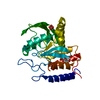
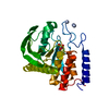
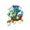
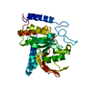
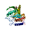
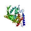
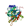
- PDB-3blt: Crystal structures of YopH complexed with PVSN and PVS, inhibitor... -
+ Open data

- Basic information
| Entry | Database: PDB / ID: 3blt | ||||||
|---|---|---|---|---|---|---|---|
| Title | Crystal structures of YopH complexed with PVSN and PVS, inhibitors of YopH which co-valent bind to Cys of active site | ||||||
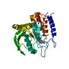
 Components Components | Tyrosine-protein phosphatase yopH | ||||||
 Keywords Keywords | HYDROLASE / Co-valent binding / binding affinity / binding selectivity / Membrane / Outer membrane / Plasmid / Protein phosphatase / Secreted / Virulence | ||||||
| Function / homology |  Function and homology information Function and homology informationprotein-tyrosine-phosphatase / protein tyrosine phosphatase activity / extracellular region Similarity search - Function | ||||||
| Biological species |  Yersinia enterocolitica (bacteria) Yersinia enterocolitica (bacteria) | ||||||
| Method |  X-RAY DIFFRACTION / FOURIER SYNTHESIS / Resolution: 2.2 Å X-RAY DIFFRACTION / FOURIER SYNTHESIS / Resolution: 2.2 Å | ||||||
 Authors Authors | Liu, S. / Zhang, Z.-Y. | ||||||
 Citation Citation | Journal: J.Am.Chem.Soc. / Year: 2008 Title: Aryl vinyl sulfonates and sulfones as active site-directed and mechanism-based probes for protein tyrosine phosphatases. Authors: Liu, S. / Zhou, B. / Yang, H. / He, Y. / Jiang, Z.X. / Kumar, S. / Wu, L. / Zhang, Z.Y. | ||||||
| History |
|
- Structure visualization
| Structure viewer | Molecule: MolmilJmol/JSmol |
|---|
- Downloads & links
-Download
| PDBx/mmCIF format | 3blt.cif.gz | 69.3 KB | Display | PDBx/mmCIF format |
|---|---|---|---|---|
| PDB format | pdb3blt.ent.gz | 50.3 KB | Display | PDB format |
| PDBx/mmJSON format | 3blt.json.gz | Tree view | PDBx/mmJSON format | |
| Others |  Other downloads Other downloads |
-Validation report
| Arichive directory | https://data.pdbj.org/pub/pdb/validation_reports/bl/3bltftp://data.pdbj.org/pub/pdb/validation_reports/bl/3blt | HTTPS FTP |
|---|
-Related structure data
| Related structure data | 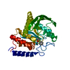 3bluC  3bm8C  1pa9S C: citing same article ( S: Starting model for refinement |
|---|---|
| Similar structure data |


-Links
 PDBj
PDBj


- Assembly
| Deposited unit | 
| ||||||||
|---|---|---|---|---|---|---|---|---|---|
| 1 |
| ||||||||
| Unit cell |
|
-Components
| #1: Protein | Mass: 33422.688 Da / Num. of mol.: 1 / Fragment: YopH catalytic domain Source method: isolated from a genetically manipulated source Source: (gene. exp.) Yersinia enterocolitica (bacteria) / Gene: yopH, yop51 / Production host: |
|---|---|
| #2: Chemical | ChemComp-PSY / 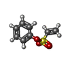  Mass: 184.212 Da / Num. of mol.: 1 / Source method: obtained synthetically / Formula: C8H8O3S Mass: 184.212 Da / Num. of mol.: 1 / Source method: obtained synthetically / Formula: C8H8O3S |
| #3: Water | ChemComp-HOH /  Mass: 18.015 Da / Num. of mol.: 94 / Source method: isolated from a natural source / Formula: H2O Mass: 18.015 Da / Num. of mol.: 94 / Source method: isolated from a natural source / Formula: H2O |
| Has protein modification | Y |
-Experimental details
-Experiment
| Experiment | Method: X-RAY DIFFRACTION / Number of used crystals: 1 |
|---|
- Sample preparation
| Crystal | Density Matthews: 2 Å3/Da / Density % sol: 38.43 % |
|---|---|
| Crystal grow | Temperature: 298 K / Method: vapor diffusion, hanging drop / pH: 7.5 Details: 25% w/v poly(ethylene glycol) 3350, 100 mM NaCl ,and 100 mM HEPES buffer (pH7.5). , VAPOR DIFFUSION, HANGING DROP, temperature 298K |
-Data collection
| Diffraction | Mean temperature: 100 K |
|---|---|
| Diffraction source | Source: ROTATING ANODE / Type: RIGAKU / Wavelength: 1.5418 Å |
| Detector | Type: RIGAKU RAXIS IV++ / Detector: IMAGE PLATE |
| Radiation | Protocol: SINGLE WAVELENGTH / Monochromatic (M) / Laue (L): M / Scattering type: x-ray |
| Radiation wavelength | Wavelength: 1.5418 Å / Relative weight: 1 |
| Reflection | Resolution: 2→50 Å / Num. obs: 16695 / % possible obs: 92.1 % / Redundancy: 3.5 % / Rmerge(I) obs: 0.047 / Net I/σ(I): 15.2 |
- Processing
| Software |
| ||||||||||||||||
|---|---|---|---|---|---|---|---|---|---|---|---|---|---|---|---|---|---|
| Refinement | Method to determine structure: FOURIER SYNTHESIS Starting model: 1pa9 Resolution: 2.2→50 Å / Cross valid method: THROUGHOUT / σ(F): 2 / Stereochemistry target values: Engh & Huber
| ||||||||||||||||
| Refinement step | Cycle: LAST / Resolution: 2.2→50 Å
| ||||||||||||||||
| Refine LS restraints |
|
