[English] 日本語
Yorodumi
- PDB-4ag2: Human Chymase - Fynomer Complex -

+
Open data


ID or keywords:

Loading...

-
Basic information

Entry
Database: PDB / ID: 4ag2
TitleHuman Chymase - Fynomer Complex
Components
  • CHYMASE
  • FYNOMER
KeywordsHYDROLASE/DE NOVO PROTEIN / HYDROLASE-DE NOVO PROTEIN COMPLEX / INHIBITOR / SERINE PROTEASE
Function / homology
Function and homology information


chymase / basement membrane disassembly / cytokine precursor processing / midbrain development / Activation of Matrix Metalloproteinases / extracellular matrix disassembly / angiotensin maturation / Metabolism of Angiotensinogen to Angiotensins / serine-type peptidase activity / secretory granule ...chymase / basement membrane disassembly / cytokine precursor processing / midbrain development / Activation of Matrix Metalloproteinases / extracellular matrix disassembly / angiotensin maturation / Metabolism of Angiotensinogen to Angiotensins / serine-type peptidase activity / secretory granule / cellular response to glucose stimulus / peptide binding / protein catabolic process / Signaling by SCF-KIT / cytoplasmic ribonucleoprotein granule / positive regulation of angiogenesis / regulation of inflammatory response / collagen-containing extracellular matrix / endopeptidase activity / intracellular membrane-bounded organelle / serine-type endopeptidase activity / extracellular space / extracellular region / cytoplasm / cytosol
Similarity search - Function
SH3 Domains / SH3 type barrels. / Serine proteases, trypsin family, histidine active site / Serine proteases, trypsin family, serine active site / Peptidase S1A, chymotrypsin family / Serine proteases, trypsin family, histidine active site. / Serine proteases, trypsin domain profile. / Serine proteases, trypsin family, serine active site. / Trypsin-like serine protease / Serine proteases, trypsin domain ...SH3 Domains / SH3 type barrels. / Serine proteases, trypsin family, histidine active site / Serine proteases, trypsin family, serine active site / Peptidase S1A, chymotrypsin family / Serine proteases, trypsin family, histidine active site. / Serine proteases, trypsin domain profile. / Serine proteases, trypsin family, serine active site. / Trypsin-like serine protease / Serine proteases, trypsin domain / Trypsin / Trypsin-like serine proteases / Thrombin, subunit H / Roll / Peptidase S1, PA clan, chymotrypsin-like fold / Peptidase S1, PA clan / Beta Barrel / Mainly Beta
Similarity search - Domain/homology
(2S)-2-hydroxybutanedioic acid / Chymase
Similarity search - Component
Biological speciesHOMO SAPIENS (human)
SYNTHETIC CONSTRUCT (others)
MethodX-RAY DIFFRACTION / SYNCHROTRON / MOLECULAR REPLACEMENT / Resolution: 1.8 Å
AuthorsSchlatter, D. / Brack, S. / Banner, D.W. / Batey, S. / Benz, J. / Bertschinger, J. / Huber, W. / Joseph, C. / Rufer, A. / Van Der Kloosters, A. ...Schlatter, D. / Brack, S. / Banner, D.W. / Batey, S. / Benz, J. / Bertschinger, J. / Huber, W. / Joseph, C. / Rufer, A. / Van Der Kloosters, A. / Weber, M. / Grabulovski, D. / Hennig, M.
CitationJournal: Mabs / Year: 2012
Title: Generation, Characterization and Structural Data of Chymase Binding Proteins Based on the Human Fyn Kinase SH3 Domain.
Authors: Schlatter, D. / Brack, S. / Banner, D.W. / Batey, S. / Benz, J. / Bertschinger, J. / Huber, W. / Joseph, C. / Rufer, A. / Van Der Klooster, A. / Weber, M. / Grabulovski, D. / Hennig, M.
History
DepositionJan 23, 2012Deposition site: PDBE / Processing site: PDBE
Revision 1.0Jul 11, 2012Provider: repository / Type: Initial release
Revision 1.1Aug 15, 2012Group: Database references / Structure summary
Revision 1.2May 1, 2024Group: Data collection / Database references ...Data collection / Database references / Derived calculations / Other / Refinement description
Category: chem_comp_atom / chem_comp_bond ...chem_comp_atom / chem_comp_bond / database_2 / pdbx_database_status / pdbx_initial_refinement_model / pdbx_struct_conn_angle / struct_conn / struct_site
Item: _database_2.pdbx_DOI / _database_2.pdbx_database_accession ..._database_2.pdbx_DOI / _database_2.pdbx_database_accession / _pdbx_database_status.status_code_sf / _pdbx_struct_conn_angle.ptnr1_auth_asym_id / _pdbx_struct_conn_angle.ptnr1_auth_comp_id / _pdbx_struct_conn_angle.ptnr1_auth_seq_id / _pdbx_struct_conn_angle.ptnr1_label_asym_id / _pdbx_struct_conn_angle.ptnr1_label_atom_id / _pdbx_struct_conn_angle.ptnr1_label_comp_id / _pdbx_struct_conn_angle.ptnr1_label_seq_id / _pdbx_struct_conn_angle.ptnr1_symmetry / _pdbx_struct_conn_angle.ptnr2_auth_seq_id / _pdbx_struct_conn_angle.ptnr2_label_asym_id / _pdbx_struct_conn_angle.ptnr3_auth_asym_id / _pdbx_struct_conn_angle.ptnr3_auth_comp_id / _pdbx_struct_conn_angle.ptnr3_auth_seq_id / _pdbx_struct_conn_angle.ptnr3_label_asym_id / _pdbx_struct_conn_angle.ptnr3_label_atom_id / _pdbx_struct_conn_angle.ptnr3_label_comp_id / _pdbx_struct_conn_angle.ptnr3_label_seq_id / _pdbx_struct_conn_angle.ptnr3_symmetry / _pdbx_struct_conn_angle.value / _struct_conn.pdbx_dist_value / _struct_conn.ptnr1_auth_asym_id / _struct_conn.ptnr1_auth_comp_id / _struct_conn.ptnr1_auth_seq_id / _struct_conn.ptnr1_label_asym_id / _struct_conn.ptnr1_label_atom_id / _struct_conn.ptnr1_label_comp_id / _struct_conn.ptnr1_label_seq_id / _struct_conn.ptnr1_symmetry / _struct_conn.ptnr2_auth_asym_id / _struct_conn.ptnr2_auth_comp_id / _struct_conn.ptnr2_auth_seq_id / _struct_conn.ptnr2_label_asym_id / _struct_conn.ptnr2_label_atom_id / _struct_conn.ptnr2_label_comp_id / _struct_conn.ptnr2_label_seq_id / _struct_conn.ptnr2_symmetry / _struct_site.pdbx_auth_asym_id / _struct_site.pdbx_auth_comp_id / _struct_site.pdbx_auth_seq_id
Remark 700 
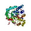
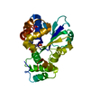
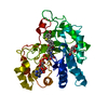
SHEET DETERMINATION METHOD: DSSP THE SHEETS PRESENTED AS "AA" IN EACH CHAIN ON SHEET RECORDS BELOW ... SHEET DETERMINATION METHOD: DSSP THE SHEETS PRESENTED AS "AA" IN EACH CHAIN ON SHEET RECORDS BELOW IS ACTUALLY AN 7-STRANDED BARREL THIS IS REPRESENTED BY A 8-STRANDED SHEET IN WHICH THE FIRST AND LAST STRANDS ARE IDENTICAL. THE SHEETS PRESENTED AS "AB" IN EACH CHAIN ON SHEET RECORDS BELOW IS ACTUALLY AN 6-STRANDED BARREL THIS IS REPRESENTED BY A 7-STRANDED SHEET IN WHICH THE FIRST AND LAST STRANDS ARE IDENTICAL. THE SHEETS PRESENTED AS "BA" IN EACH CHAIN ON SHEET RECORDS BELOW IS ACTUALLY AN 7-STRANDED BARREL THIS IS REPRESENTED BY A 8-STRANDED SHEET IN WHICH THE FIRST AND LAST STRANDS ARE IDENTICAL. THE SHEETS PRESENTED AS "BB" IN EACH CHAIN ON SHEET RECORDS BELOW IS ACTUALLY AN 6-STRANDED BARREL THIS IS REPRESENTED BY A 7-STRANDED SHEET IN WHICH THE FIRST AND LAST STRANDS ARE IDENTICAL.

-
Structure visualization

Structure viewerMolecule:
MolmilJmol/JSmol

Downloads & links

-
Assembly

Deposited unit
A: CHYMASE
B: CHYMASE
C: FYNOMER
D: FYNOMER
hetero molecules


Theoretical massNumber of molelcules
Total (without water)70,26914
Polymers69,3614
Non-polymers90810
Water9,260514
1
A: CHYMASE
C: FYNOMER
hetero molecules


Theoretical massNumber of molelcules
Total (without water)35,2979
Polymers34,6802
Non-polymers6177
Water362
TypeNameSymmetry operationNumber
identity operation1_555x,y,z1
Buried area2440 Å2
ΔGint-20.7 kcal/mol
Surface area12920 Å2
MethodPISA
2
B: CHYMASE
D: FYNOMER
hetero molecules


Theoretical massNumber of molelcules
Total (without water)34,9725
Polymers34,6802
Non-polymers2913
Water362
TypeNameSymmetry operationNumber
identity operation1_555x,y,z1
Buried area2360 Å2
ΔGint-13.1 kcal/mol
Surface area12880 Å2
MethodPISA
Unit cell
Length a, b, c (Å)56.937, 64.124, 174.987
Angle α, β, γ (deg.)90.00, 90.00, 90.00
Int Tables number19
Space group name H-MP212121

-
Components

-
Protein , 2 types, 4 molecules ABCD

#1: Protein CHYMASE / ALPHA-CHYMASE / MAST CELL PROTEASE I


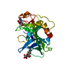
Mass: 25066.969 Da / Num. of mol.: 2
Source method: isolated from a genetically manipulated source
Source: (gene. exp.) HOMO SAPIENS (human) / Production host: ESCHERICHIA COLI (E. coli) / References: UniProt: P23946, chymase
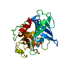
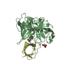
#2: Protein FYNOMER


Mass: 9613.473 Da / Num. of mol.: 2
Source method: isolated from a genetically manipulated source
Source: (gene. exp.) SYNTHETIC CONSTRUCT (others) / Production host: ESCHERICHIA COLI (E. coli)

-
Non-polymers , 4 types, 524 molecules

#3: Chemical ChemComp-MES / 2-(N-MORPHOLINO)-ETHANESULFONIC ACID


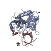
Mass: 195.237 Da / Num. of mol.: 2 / Source method: obtained synthetically / Formula: C6H13NO4S / Comment: pH buffer*YM
#4: Chemical ChemComp-LMR / (2S)-2-hydroxybutanedioic acid / L-Malate


Mass: 134.087 Da / Num. of mol.: 3 / Source method: obtained synthetically / Formula: C4H6O5
#5: Chemical
ChemComp-NA / SODIUM ION


Mass: 22.990 Da / Num. of mol.: 5 / Source method: obtained synthetically / Formula: Na
#6: Water ChemComp-HOH / water


Mass: 18.015 Da / Num. of mol.: 514 / Source method: isolated from a natural source / Formula: H2O

-
Details

Sequence detailsCHAINS C, D: ARTIFICIAL PROTEIN BASED ON SH3 DOMAIN OF P06241 (83-145)

-
Experimental details

-
Experiment

ExperimentMethod: X-RAY DIFFRACTION / Number of used crystals: 1

-
Sample preparation

CrystalDensity Matthews: 2.55 Å3/Da / Density % sol: 52 % / Description: NONE
Crystal growpH: 7 / Details: 0.15 M DL-MALIC ACID PH 7.0, 20% PEG 3350

-
Data collection

DiffractionMean temperature: 100 K
Diffraction sourceSource: SYNCHROTRON / Site: SLS / Beamline: X10SA / Wavelength: 0.9999
DetectorType: DECTRIS PILATUS 6M / Detector: PIXEL / Date: Jun 2, 2010
RadiationProtocol: SINGLE WAVELENGTH / Monochromatic (M) / Laue (L): M / Scattering type: x-ray
Radiation wavelengthWavelength: 0.9999 Å / Relative weight: 1
ReflectionResolution: 1.78→50 Å / Num. obs: 62210 / % possible obs: 99.7 % / Observed criterion σ(I): -3 / Redundancy: 6.19 % / Biso Wilson estimate: 32 Å2 / Rmerge(I) obs: 0.09 / Net I/σ(I): 10.95
Reflection shellResolution: 1.78→1.87 Å / Redundancy: 5.62 % / Rmerge(I) obs: 0.67 / Mean I/σ(I) obs: 1.49 / % possible all: 99

-
Processing

Software
NameVersionClassification
REFMAC5.6.0070refinement
XDSdata reduction
SADABSdata scaling
PHASERphasing
RefinementMethod to determine structure: MOLECULAR REPLACEMENT
Starting model: IN HOUSE STRUCTURE

Resolution: 1.8→43.75 Å / Cor.coef. Fo:Fc: 0.964 / Cor.coef. Fo:Fc free: 0.95 / SU B: 2.92 / SU ML: 0.087 / Cross valid method: THROUGHOUT / ESU R: 0.125 / ESU R Free: 0.118 / Stereochemistry target values: MAXIMUM LIKELIHOOD
Details: HYDROGENS HAVE BEEN ADDED IN THE RIDING POSITIONS. HYDROGENS USED BUT NOT OUTPUT.
RfactorNum. reflection% reflectionSelection details
Rfree0.21168 2917 5 %RANDOM
Rwork0.17853 ---
obs0.18021 55464 96.75 %-
Solvent computationIon probe radii: 0.8 Å / Shrinkage radii: 0.8 Å / VDW probe radii: 1.2 Å / Solvent model: BABINET MODEL WITH MASK
Displacement parametersBiso mean: 28.461 Å2
Baniso -1Baniso -2Baniso -3
1-0.08 Å20 Å20 Å2
2--0.78 Å20 Å2
3----0.86 Å2
Refinement stepCycle: LAST / Resolution: 1.8→43.75 Å
ProteinNucleic acidLigandSolventTotal
Num. atoms4435 0 56 514 5005
Refine LS restraints
Refine-IDTypeDev idealDev ideal targetNumber
X-RAY DIFFRACTIONr_bond_refined_d0.0120.0224662
X-RAY DIFFRACTIONr_bond_other_d
X-RAY DIFFRACTIONr_angle_refined_deg1.3561.9556339
X-RAY DIFFRACTIONr_angle_other_deg
X-RAY DIFFRACTIONr_dihedral_angle_1_deg6.3775570
X-RAY DIFFRACTIONr_dihedral_angle_2_deg32.51422.921202
X-RAY DIFFRACTIONr_dihedral_angle_3_deg12.82515745
X-RAY DIFFRACTIONr_dihedral_angle_4_deg11.0291532
X-RAY DIFFRACTIONr_chiral_restr0.0960.2683
X-RAY DIFFRACTIONr_gen_planes_refined0.0060.0213565
X-RAY DIFFRACTIONr_gen_planes_other
X-RAY DIFFRACTIONr_nbd_refined
X-RAY DIFFRACTIONr_nbd_other
X-RAY DIFFRACTIONr_nbtor_refined
X-RAY DIFFRACTIONr_nbtor_other
X-RAY DIFFRACTIONr_xyhbond_nbd_refined
X-RAY DIFFRACTIONr_xyhbond_nbd_other
X-RAY DIFFRACTIONr_metal_ion_refined
X-RAY DIFFRACTIONr_metal_ion_other
X-RAY DIFFRACTIONr_symmetry_vdw_refined
X-RAY DIFFRACTIONr_symmetry_vdw_other
X-RAY DIFFRACTIONr_symmetry_hbond_refined
X-RAY DIFFRACTIONr_symmetry_hbond_other
X-RAY DIFFRACTIONr_symmetry_metal_ion_refined
X-RAY DIFFRACTIONr_symmetry_metal_ion_other
X-RAY DIFFRACTIONr_mcbond_it
X-RAY DIFFRACTIONr_mcbond_other
X-RAY DIFFRACTIONr_mcangle_it
X-RAY DIFFRACTIONr_mcangle_other
X-RAY DIFFRACTIONr_scbond_it
X-RAY DIFFRACTIONr_scbond_other
X-RAY DIFFRACTIONr_scangle_it
X-RAY DIFFRACTIONr_scangle_other
X-RAY DIFFRACTIONr_long_range_B_refined
X-RAY DIFFRACTIONr_long_range_B_other
X-RAY DIFFRACTIONr_rigid_bond_restr
X-RAY DIFFRACTIONr_sphericity_free
X-RAY DIFFRACTIONr_sphericity_bonded
LS refinement shellResolution: 1.8→1.847 Å / Total num. of bins used: 20
RfactorNum. reflection% reflection
Rfree0.325 198 -
Rwork0.32 3763 -
obs--89.74 %

+
About Yorodumi

-
News

-
Feb 9, 2022. New format data for meta-information of EMDB entries

New format data for meta-information of EMDB entries

  • Version 3 of the EMDB header file is now the official format.
  • The previous official version 1.9 will be removed from the archive.

Related info.:EMDB header

External links:wwPDB to switch to version 3 of the EMDB data model

-
Aug 12, 2020. Covid-19 info

Covid-19 info

URL: https://pdbjlvh1.pdbj.org/emnavi/covid19.php

New page: Covid-19 featured information page in EM Navigator.

Related info.:Covid-19 info / Mar 5, 2020. Novel coronavirus structure data

+
Mar 5, 2020. Novel coronavirus structure data

Novel coronavirus structure data

Related info.:Yorodumi Speices / Aug 12, 2020. Covid-19 info

External links:COVID-19 featured content - PDBj / Molecule of the Month (242):Coronavirus Proteases

+
Jan 31, 2019. EMDB accession codes are about to change! (news from PDBe EMDB page)

EMDB accession codes are about to change! (news from PDBe EMDB page)

  • The allocation of 4 digits for EMDB accession codes will soon come to an end. Whilst these codes will remain in use, new EMDB accession codes will include an additional digit and will expand incrementally as the available range of codes is exhausted. The current 4-digit format prefixed with “EMD-” (i.e. EMD-XXXX) will advance to a 5-digit format (i.e. EMD-XXXXX), and so on. It is currently estimated that the 4-digit codes will be depleted around Spring 2019, at which point the 5-digit format will come into force.
  • The EM Navigator/Yorodumi systems omit the EMD- prefix.

Related info.:Q: What is EMD? / ID/Accession-code notation in Yorodumi/EM Navigator

External links:EMDB Accession Codes are Changing Soon! / Contact to PDBj

+
Jul 12, 2017. Major update of PDB

Major update of PDB

  • wwPDB released updated PDB data conforming to the new PDBx/mmCIF dictionary.
  • This is a major update changing the version number from 4 to 5, and with Remediation, in which all the entries are updated.
  • In this update, many items about electron microscopy experimental information are reorganized (e.g. em_software).
  • Now, EM Navigator and Yorodumi are based on the updated data.

External links:wwPDB Remediation / Enriched Model Files Conforming to OneDep Data Standards Now Available in the PDB FTP Archive

-
Yorodumi

Thousand views of thousand structures

  • Yorodumi is a browser for structure data from EMDB, PDB, SASBDB, etc.
  • This page is also the successor to EM Navigator detail page, and also detail information page/front-end page for Omokage search.
  • The word "yorodu" (or yorozu) is an old Japanese word meaning "ten thousand". "mi" (miru) is to see.

Related info.:EMDB / PDB / SASBDB / Comparison of 3 databanks / Yorodumi Search / Aug 31, 2016. New EM Navigator & Yorodumi / Yorodumi Papers / Jmol/JSmol / Function and homology information / Changes in new EM Navigator and Yorodumi

Read more