 Movie
Movie Controller

[English] 日本語
 Yorodumi
- PDB-1klt: CRYSTAL STRUCTURE OF PMSF-TREATED HUMAN CHYMASE AT 1.9 ANGSTROMS ... -
+ Open data

- Basic information
| Entry | Database: PDB / ID: 1klt | ||||||
|---|---|---|---|---|---|---|---|
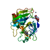
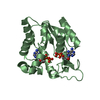
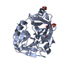
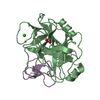
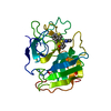
| Title | CRYSTAL STRUCTURE OF PMSF-TREATED HUMAN CHYMASE AT 1.9 ANGSTROMS RESOLUTION | ||||||
 Components Components | CHYMASE | ||||||
 Keywords Keywords | SERINE PROTEASE / HYDROLASE / MAST CELL / ANGIOTENSIN / ALPHA TOLUENESULFONIC ACID | ||||||
| Function / homology |  Function and homology information Function and homology informationchymase / basement membrane disassembly / cytokine precursor processing / peptide metabolic process / midbrain development / Activation of Matrix Metalloproteinases / extracellular matrix disassembly / Metabolism of Angiotensinogen to Angiotensins / angiotensin maturation / peptide binding ...chymase / basement membrane disassembly / cytokine precursor processing / peptide metabolic process / midbrain development / Activation of Matrix Metalloproteinases / extracellular matrix disassembly / Metabolism of Angiotensinogen to Angiotensins / angiotensin maturation / peptide binding / secretory granule / serine-type peptidase activity / protein catabolic process / cellular response to glucose stimulus / protein maturation / Signaling by SCF-KIT / positive regulation of angiogenesis / cytoplasmic ribonucleoprotein granule / extracellular matrix / regulation of inflammatory response / endopeptidase activity / serine-type endopeptidase activity / : / extracellular region Similarity search - Function | ||||||
| Biological species |  Homo sapiens (human) Homo sapiens (human) | ||||||
| Method |  X-RAY DIFFRACTION / MOLECULAR REPLACEMENT / Resolution: 1.9 Å X-RAY DIFFRACTION / MOLECULAR REPLACEMENT / Resolution: 1.9 Å | ||||||
 Authors Authors | Mcgrath, M.E. | ||||||
 Citation Citation | Journal: Biochemistry / Year: 1997 Title: Crystal structure of phenylmethanesulfonyl fluoride-treated human chymase at 1.9 A. Authors: McGrath, M.E. / Mirzadegan, T. / Schmidt, B.F. #1: Journal: FEBS Lett. / Year: 1997Title: Production of Crystallizable Human Chymase from a Bacillus Subtilis System Authors: Mcgrath, M.E. / Osawa, A.E. / Barnes, M.G. / Clark, J.M. / Mortara, K.D. / Schmidt, B.F. | ||||||
| History |
|
- Structure visualization
| Structure viewer | Molecule: MolmilJmol/JSmol |
|---|
- Downloads & links
-Download
| PDBx/mmCIF format | 1klt.cif.gz | 79.7 KB | Display | PDBx/mmCIF format |
|---|---|---|---|---|
| PDB format | pdb1klt.ent.gz | 58.7 KB | Display | PDB format |
| PDBx/mmJSON format | 1klt.json.gz | Tree view | PDBx/mmJSON format | |
| Others |  Other downloads Other downloads |
-Validation report
| Arichive directory | https://data.pdbj.org/pub/pdb/validation_reports/kl/1kltftp://data.pdbj.org/pub/pdb/validation_reports/kl/1klt | HTTPS FTP |
|---|
-Related structure data
| Similar structure data |
|---|


-Links
 PDBj
PDBj


- Assembly
| Deposited unit | 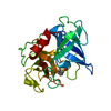
| ||||||||
|---|---|---|---|---|---|---|---|---|---|
| 1 |
| ||||||||
| Unit cell |
|
-Components
| #1: Protein | Mass: 25050.904 Da / Num. of mol.: 1 / Mutation: C22S Source method: isolated from a genetically manipulated source Source: (gene. exp.) Homo sapiens (human) / Production host:  |
|---|---|
| #2: Chemical | ChemComp-PMS /   Mass: 172.202 Da / Num. of mol.: 1 / Source method: obtained synthetically / Formula: C7H8O3S Mass: 172.202 Da / Num. of mol.: 1 / Source method: obtained synthetically / Formula: C7H8O3S |
| #3: Water | ChemComp-HOH /  Mass: 18.015 Da / Num. of mol.: 221 / Source method: isolated from a natural source / Formula: H2O Mass: 18.015 Da / Num. of mol.: 221 / Source method: isolated from a natural source / Formula: H2O |
| Has protein modification | Y |
-Experimental details
-Experiment
| Experiment | Method: X-RAY DIFFRACTION / Number of used crystals: 1 |
|---|
- Sample preparation
| Crystal | Density Matthews: 2.3 Å3/Da / Density % sol: 46.5 % | |||||||||||||||||||||||||
|---|---|---|---|---|---|---|---|---|---|---|---|---|---|---|---|---|---|---|---|---|---|---|---|---|---|---|
| Crystal grow | pH: 5.6 Details: COMPLEX WAS CRYSTALLIZED FROM 20% PEG 2000, 20% ISOPROPANOL, 0.1M SODIUM CITRATE, PH 5.6. | |||||||||||||||||||||||||
| Crystal grow | *PLUS Method: vapor diffusion, hanging drop | |||||||||||||||||||||||||
| Components of the solutions | *PLUS
|
-Data collection
| Diffraction | Mean temperature: 295 K |
|---|---|
| Diffraction source | Source: ROTATING ANODE / Type: RIGAKU RUH2R / Wavelength: 1.5418 |
| Detector | Type: RIGAKU / Detector: IMAGE PLATE / Date: Dec 1, 1996 / Details: MIRRORS |
| Radiation | Monochromator: NI FILTER / Monochromatic (M) / Laue (L): M / Scattering type: x-ray |
| Radiation wavelength | Wavelength: 1.5418 Å / Relative weight: 1 |
| Reflection | Resolution: 1.9→39.1 Å / Num. obs: 14518 / % possible obs: 80.4 % / Observed criterion σ(I): 1 / Redundancy: 2.2 % / Biso Wilson estimate: 21.3 Å2 / Rmerge(I) obs: 0.077 / Rsym value: 0.077 / Net I/σ(I): 11 |
| Reflection shell | Resolution: 1.9→2.1 Å / Rmerge(I) obs: 0.344 / Mean I/σ(I) obs: 1.3 / Rsym value: 0.344 / % possible all: 50.4 |
| Reflection | *PLUS Num. measured all: 31632 |
| Reflection shell | *PLUS % possible obs: 50.4 % |
- Processing
| Software |
| ||||||||||||||||||||||||||||||||||||||||||||||||||||||||||||
|---|---|---|---|---|---|---|---|---|---|---|---|---|---|---|---|---|---|---|---|---|---|---|---|---|---|---|---|---|---|---|---|---|---|---|---|---|---|---|---|---|---|---|---|---|---|---|---|---|---|---|---|---|---|---|---|---|---|---|---|---|---|
| Refinement | Method to determine structure: MOLECULAR REPLACEMENT Starting model: HOMOLOGY MODEL Resolution: 1.9→6 Å / Rfactor Rfree error: 0.017 / Data cutoff high absF: 10000000 / Data cutoff low absF: 0.001 / Cross valid method: THROUGHOUT / σ(F): 1
| ||||||||||||||||||||||||||||||||||||||||||||||||||||||||||||
| Displacement parameters | Biso mean: 19.4 Å2 | ||||||||||||||||||||||||||||||||||||||||||||||||||||||||||||
| Refine analyze | Luzzati coordinate error obs: 0.21 Å / Luzzati d res low obs: 6 Å | ||||||||||||||||||||||||||||||||||||||||||||||||||||||||||||
| Refinement step | Cycle: LAST / Resolution: 1.9→6 Å
| ||||||||||||||||||||||||||||||||||||||||||||||||||||||||||||
| Refine LS restraints |
| ||||||||||||||||||||||||||||||||||||||||||||||||||||||||||||
| LS refinement shell | Resolution: 1.9→1.98 Å / Rfactor Rfree error: 0.042 / Total num. of bins used: 8
| ||||||||||||||||||||||||||||||||||||||||||||||||||||||||||||
| Xplor file |
| ||||||||||||||||||||||||||||||||||||||||||||||||||||||||||||
| Software | *PLUS Name: X-PLOR / Version: 3.1 / Classification: refinement | ||||||||||||||||||||||||||||||||||||||||||||||||||||||||||||
| Refinement | *PLUS | ||||||||||||||||||||||||||||||||||||||||||||||||||||||||||||
| Solvent computation | *PLUS | ||||||||||||||||||||||||||||||||||||||||||||||||||||||||||||
| Displacement parameters | *PLUS | ||||||||||||||||||||||||||||||||||||||||||||||||||||||||||||
| Refine LS restraints | *PLUS
| ||||||||||||||||||||||||||||||||||||||||||||||||||||||||||||
| LS refinement shell | *PLUS Rfactor obs: 0.321 |
