 Movie
Movie Controller


+ Open data

- Basic information
| Entry | Database: PDB / ID: 3lz5 | ||||||
|---|---|---|---|---|---|---|---|
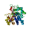
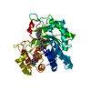
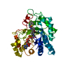
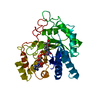
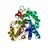
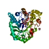
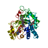
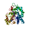
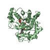
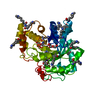
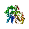
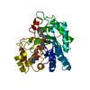
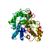
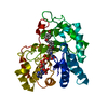
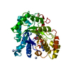
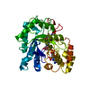
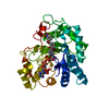
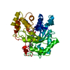
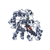
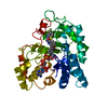
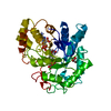
| Title | Human aldose reductase mutant T113V complexed with IDD594 | ||||||
 Components Components | Aldose Reductase | ||||||
 Keywords Keywords | OXIDOREDUCTASE/OXIDOREDUCTASE INHIBITOR / T113V mutant / oxidoreductase / TIM barrel / NADP / Phosphoprotein / OXIDOREDUCTASE-OXIDOREDUCTASE INHIBITOR complex | ||||||
| Function / homology |  Function and homology information Function and homology informationglyceraldehyde oxidoreductase activity / Fructose biosynthesis / fructose biosynthetic process / L-glucuronate reductase activity / aldose reductase / D/L-glyceraldehyde reductase / glycerol dehydrogenase (NADP+) activity / C21-steroid hormone biosynthetic process / NADP-retinol dehydrogenase / Pregnenolone biosynthesis ...glyceraldehyde oxidoreductase activity / Fructose biosynthesis / fructose biosynthetic process / L-glucuronate reductase activity / aldose reductase / D/L-glyceraldehyde reductase / glycerol dehydrogenase (NADP+) activity / C21-steroid hormone biosynthetic process / NADP-retinol dehydrogenase / Pregnenolone biosynthesis / allyl-alcohol dehydrogenase / allyl-alcohol dehydrogenase activity / Galactose catabolism / prostaglandin H2 endoperoxidase reductase activity / regulation of urine volume / all-trans-retinol dehydrogenase (NADP+) activity / metanephric collecting duct development / daunorubicin metabolic process / doxorubicin metabolic process / retinal dehydrogenase (NAD+) activity / epithelial cell maturation / aldose reductase (NADPH) activity / cellular hyperosmotic salinity response / renal water homeostasis / retinoid metabolic process / carbohydrate metabolic process / electron transfer activity / negative regulation of apoptotic process / mitochondrion / : / extracellular exosome / nucleoplasm / cytosol Similarity search - Function | ||||||
| Biological species |  Homo sapiens (human) Homo sapiens (human) | ||||||
| Method |  X-RAY DIFFRACTION / SYNCHROTRON / AB INITIO PHASING / Resolution: 0.95 Å X-RAY DIFFRACTION / SYNCHROTRON / AB INITIO PHASING / Resolution: 0.95 Å | ||||||
 Authors Authors | Koch, C. / Heine, A. / Klebe, G. | ||||||
 Citation Citation | Journal: J.Mol.Biol. / Year: 2011 Title: Tracing the detail: how mutations affect binding modes and thermodynamic signatures of closely related aldose reductase inhibitors Authors: Koch, C. / Heine, A. / Klebe, G. | ||||||
| History |
|
- Structure visualization
| Structure viewer | Molecule: MolmilJmol/JSmol |
|---|
- Downloads & links
-Download
| PDBx/mmCIF format | 3lz5.cif.gz | 161.7 KB | Display | PDBx/mmCIF format |
|---|---|---|---|---|
| PDB format | pdb3lz5.ent.gz | 125.4 KB | Display | PDB format |
| PDBx/mmJSON format | 3lz5.json.gz | Tree view | PDBx/mmJSON format | |
| Others |  Other downloads Other downloads |
-Validation report
| Arichive directory | https://data.pdbj.org/pub/pdb/validation_reports/lz/3lz5ftp://data.pdbj.org/pub/pdb/validation_reports/lz/3lz5 | HTTPS FTP |
|---|
-Related structure data
| Related structure data | 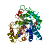 3lboC 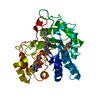 3ld5C 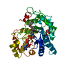 3lepC  3lqgC 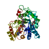 3lqlC 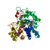 3lz3C 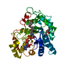 3m4hC C: citing same article ( |
|---|---|
| Similar structure data |

-Links
 PDBj
PDBj
- Assembly
| Deposited unit | 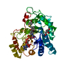
| ||||||||
|---|---|---|---|---|---|---|---|---|---|
| 1 |
| ||||||||
| Unit cell |
|
-Components
| #1: Protein | Mass: 35896.367 Da / Num. of mol.: 1 / Mutation: T113V Source method: isolated from a genetically manipulated source Source: (gene. exp.) Homo sapiens (human) / Gene: alr2 / Plasmid: pET15b / Production host:  |
|---|---|
| #2: Chemical | ChemComp-NAP /   Mass: 743.405 Da / Num. of mol.: 1 / Source method: obtained synthetically / Formula: C21H28N7O17P3 Mass: 743.405 Da / Num. of mol.: 1 / Source method: obtained synthetically / Formula: C21H28N7O17P3 |
| #3: Chemical | ChemComp-LDT /   Mass: 416.237 Da / Num. of mol.: 1 / Source method: obtained synthetically / Formula: C16H12BrF2NO3S Mass: 416.237 Da / Num. of mol.: 1 / Source method: obtained synthetically / Formula: C16H12BrF2NO3S |
| #4: Chemical | ChemComp-CIT /   Mass: 192.124 Da / Num. of mol.: 1 / Source method: obtained synthetically / Formula: C6H8O7 Mass: 192.124 Da / Num. of mol.: 1 / Source method: obtained synthetically / Formula: C6H8O7 |
| #5: Water | ChemComp-HOH /  Mass: 18.015 Da / Num. of mol.: 413 / Source method: isolated from a natural source / Formula: H2O Mass: 18.015 Da / Num. of mol.: 413 / Source method: isolated from a natural source / Formula: H2O |
| Sequence details | THIS SEQUENCE CONFLICT HAS BEEN DESCRIBED IN J BIOL CHEM 1989 SEP 5; 264(25): 14775-7 BY CHUNG S, ...THIS SEQUENCE CONFLICT HAS BEEN DESCRIBED IN J BIOL CHEM 1989 SEP 5; 264(25): 14775-7 BY CHUNG S, ET AL., PUBMED ID 2504709 |
-Experimental details
-Experiment
| Experiment | Method: X-RAY DIFFRACTION / Number of used crystals: 1 |
|---|
- Sample preparation
| Crystal | Density Matthews: 2.07 Å3/Da / Density % sol: 43.33 % |
|---|---|
| Crystal grow | Temperature: 293 K / Method: vapor diffusion, hanging drop / pH: 5 Details: 20% PEG 6000, 120mM ammonium citrate, pH 5.0, VAPOR DIFFUSION, HANGING DROP, temperature 293K |
-Data collection
| Diffraction | Mean temperature: 100 K |
|---|---|
| Diffraction source | Source: SYNCHROTRON / Site: BESSY  / Beamline: 14.2 / Wavelength: 0.91841 Å / Beamline: 14.2 / Wavelength: 0.91841 Å |
| Detector | Type: MARMOSAIC 225 mm CCD / Detector: CCD / Date: Nov 7, 2008 / Details: silicon, Rh-coated |
| Radiation | Monochromator: Si-111 crystal / Protocol: SINGLE WAVELENGTH / Monochromatic (M) / Laue (L): M / Scattering type: x-ray |
| Radiation wavelength | Wavelength: 0.91841 Å / Relative weight: 1 |
| Reflection | Resolution: 0.95→20 Å / Num. all: 184645 / Num. obs: 184645 / % possible obs: 95.9 % / Observed criterion σ(F): 0 / Observed criterion σ(I): 0 / Redundancy: 2.4 % / Rsym value: 0.043 / Net I/σ(I): 19.9 |
| Reflection shell | Resolution: 0.95→0.97 Å / Redundancy: 1.9 % / Mean I/σ(I) obs: 1.9 / Num. unique all: 7801 / Rsym value: 0.385 / % possible all: 81 |
- Processing
| Software |
| |||||||||||||||||||||||||||||||||
|---|---|---|---|---|---|---|---|---|---|---|---|---|---|---|---|---|---|---|---|---|---|---|---|---|---|---|---|---|---|---|---|---|---|---|
| Refinement | Method to determine structure: AB INITIO PHASING / Resolution: 0.95→20 Å / Num. parameters: 27818 / Num. restraintsaints: 35880 / Cross valid method: FREE R / σ(F): 0 / σ(I): 0 / Stereochemistry target values: Engh & Huber Details: ANISOTROPIC SCALING APPLIED BY THE METHOD OF PARKIN, MOEZZI & HOPE, J.APPL.CRYST.28(1995)53-56 ANISOTROPIC REFINEMENT REDUCED FREE R (NO CUTOFF) BY ?
| |||||||||||||||||||||||||||||||||
| Refine analyze | Num. disordered residues: 24 / Occupancy sum hydrogen: 2466 / Occupancy sum non hydrogen: 2946.22 | |||||||||||||||||||||||||||||||||
| Refinement step | Cycle: LAST / Resolution: 0.95→20 Å
| |||||||||||||||||||||||||||||||||
| Refine LS restraints |
|
