 Movie
Movie Controller


+ Open data

- Basic information
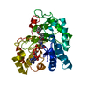
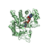
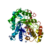
| Entry | Database: PDB / ID: 2pdc | ||||||
|---|---|---|---|---|---|---|---|
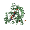
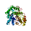
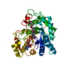
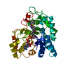
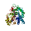
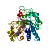
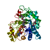
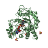
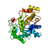
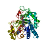
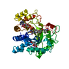
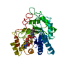
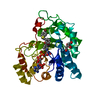
| Title | Human aldose reductase mutant F121P complexed with IDD393. | ||||||
 Components Components | Aldose reductase | ||||||
 Keywords Keywords | OXIDOREDUCTASE / TIM barrel | ||||||
| Function / homology |  Function and homology information Function and homology informationglyceraldehyde oxidoreductase activity / Fructose biosynthesis / fructose biosynthetic process / L-glucuronate reductase activity / aldose reductase / D/L-glyceraldehyde reductase / glycerol dehydrogenase (NADP+) activity / C21-steroid hormone biosynthetic process / NADP-retinol dehydrogenase / Pregnenolone biosynthesis ...glyceraldehyde oxidoreductase activity / Fructose biosynthesis / fructose biosynthetic process / L-glucuronate reductase activity / aldose reductase / D/L-glyceraldehyde reductase / glycerol dehydrogenase (NADP+) activity / C21-steroid hormone biosynthetic process / NADP-retinol dehydrogenase / Pregnenolone biosynthesis / allyl-alcohol dehydrogenase / allyl-alcohol dehydrogenase activity / Galactose catabolism / prostaglandin H2 endoperoxidase reductase activity / regulation of urine volume / all-trans-retinol dehydrogenase (NADP+) activity / metanephric collecting duct development / daunorubicin metabolic process / doxorubicin metabolic process / retinal dehydrogenase (NAD+) activity / epithelial cell maturation / aldose reductase (NADPH) activity / cellular hyperosmotic salinity response / retinoid metabolic process / renal water homeostasis / carbohydrate metabolic process / electron transfer activity / negative regulation of apoptotic process / mitochondrion / : / extracellular exosome / nucleoplasm / cytosol Similarity search - Function | ||||||
| Biological species |  Homo sapiens (human) Homo sapiens (human) | ||||||
| Method |  X-RAY DIFFRACTION / MOLECULAR REPLACEMENT / Resolution: 1.65 Å X-RAY DIFFRACTION / MOLECULAR REPLACEMENT / Resolution: 1.65 Å | ||||||
 Authors Authors | Steuber, H. / Heine, A. / Klebe, G. | ||||||
 Citation Citation | Journal: J.Mol.Biol. / Year: 2008 Title: Merging the binding sites of aldose and aldehyde reductase for detection of inhibitor selectivity-determining features. Authors: Steuber, H. / Heine, A. / Podjarny, A. / Klebe, G. | ||||||
| History |
|
- Structure visualization
| Structure viewer | Molecule: MolmilJmol/JSmol |
|---|
- Downloads & links
-Download
| PDBx/mmCIF format | 2pdc.cif.gz | 87 KB | Display | PDBx/mmCIF format |
|---|---|---|---|---|
| PDB format | pdb2pdc.ent.gz | 62.7 KB | Display | PDB format |
| PDBx/mmJSON format | 2pdc.json.gz | Tree view | PDBx/mmJSON format | |
| Others |  Other downloads Other downloads |
-Validation report
| Arichive directory | https://data.pdbj.org/pub/pdb/validation_reports/pd/2pdcftp://data.pdbj.org/pub/pdb/validation_reports/pd/2pdc | HTTPS FTP |
|---|
-Related structure data
| Related structure data | 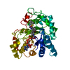 2pd5C 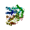 2pd9C 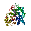 2pdbC 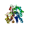 2pdfC 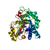 2pdgC 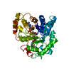 2pdhC  2pdiC 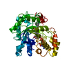 2pdjC  2pdkC 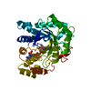 2pdlC 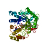 2pdmC 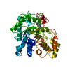 2pdnC 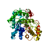 2pdpC 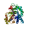 2pdqC 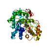 2pduC  2pdwC 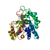 2pdxC 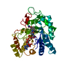 2pdyC  1el3S C: citing same article ( S: Starting model for refinement |
|---|---|
| Similar structure data |


-Links
 PDBj
PDBj
- Assembly
| Deposited unit | 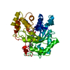
| ||||||||
|---|---|---|---|---|---|---|---|---|---|
| 1 |
| ||||||||
| Unit cell |
|
-Components
| #1: Protein | Mass: 35848.281 Da / Num. of mol.: 1 / Mutation: F121P Source method: isolated from a genetically manipulated source Source: (gene. exp.) Homo sapiens (human) / Gene: ALR2 / Plasmid: pET15 b / Production host:  |
|---|---|
| #2: Chemical | ChemComp-NAP /   Mass: 743.405 Da / Num. of mol.: 1 / Source method: obtained synthetically / Formula: C21H28N7O17P3 Mass: 743.405 Da / Num. of mol.: 1 / Source method: obtained synthetically / Formula: C21H28N7O17P3 |
| #3: Chemical | ChemComp-393 / (  Mass: 364.737 Da / Num. of mol.: 1 / Source method: obtained synthetically / Formula: C16H13ClN2O6 Mass: 364.737 Da / Num. of mol.: 1 / Source method: obtained synthetically / Formula: C16H13ClN2O6 |
| #4: Water | ChemComp-HOH /  Mass: 18.015 Da / Num. of mol.: 290 / Source method: isolated from a natural source / Formula: H2O Mass: 18.015 Da / Num. of mol.: 290 / Source method: isolated from a natural source / Formula: H2O |
-Experimental details
-Experiment
| Experiment | Method: X-RAY DIFFRACTION / Number of used crystals: 1 |
|---|
- Sample preparation
| Crystal | Density Matthews: 2.2 Å3/Da / Density % sol: 44.69 % |
|---|---|
| Crystal grow | Temperature: 293 K / Method: vapor diffusion, hanging drop / pH: 5 Details: 20 % PEG 6000 in 120 mM ammonium citrate, pH 5.0, VAPOR DIFFUSION, HANGING DROP, temperature 293K |
-Data collection
| Diffraction | Mean temperature: 100 K |
|---|---|
| Diffraction source | Source: ROTATING ANODE / Type: RIGAKU RU300 / Wavelength: 1.5418 Å |
| Detector | Type: RIGAKU RAXIS IV / Detector: IMAGE PLATE / Date: Jan 23, 2006 / Details: mirrors |
| Radiation | Monochromator: graphite / Protocol: SINGLE WAVELENGTH / Monochromatic (M) / Laue (L): M / Scattering type: x-ray |
| Radiation wavelength | Wavelength: 1.5418 Å / Relative weight: 1 |
| Reflection | Resolution: 1.65→50 Å / Num. all: 28734 / Num. obs: 28734 / % possible obs: 77.1 % / Redundancy: 1.8 % / Rmerge(I) obs: 0.02 / Rsym value: 0.02 / Net I/σ(I): 39.7 |
| Reflection shell | Resolution: 1.65→1.68 Å / Redundancy: 1.8 % / Rmerge(I) obs: 0.047 / Mean I/σ(I) obs: 19.2 / Num. unique all: 1347 / Rsym value: 0.047 / % possible all: 72.5 |
- Processing
| Software |
| |||||||||||||||||||||||||||||||||
|---|---|---|---|---|---|---|---|---|---|---|---|---|---|---|---|---|---|---|---|---|---|---|---|---|---|---|---|---|---|---|---|---|---|---|
| Refinement | Method to determine structure: MOLECULAR REPLACEMENT Starting model: 1el3 Resolution: 1.65→30 Å / Num. parameters: 11610 / Num. restraintsaints: 10770 / Cross valid method: FREE R / σ(F): 0 / σ(I): 0 / Stereochemistry target values: ENGH AND HUBER Details: ANISOTROPIC SCALING APPLIED BY THE METHOD OF PARKIN, MOEZZI & HOPE, J.APPL.CRYST.28(1995)53-56
| |||||||||||||||||||||||||||||||||
| Refine analyze | Num. disordered residues: 3 / Occupancy sum hydrogen: 2518 / Occupancy sum non hydrogen: 2886 | |||||||||||||||||||||||||||||||||
| Refinement step | Cycle: LAST / Resolution: 1.65→30 Å
| |||||||||||||||||||||||||||||||||
| Refine LS restraints |
|
