 Movie
Movie Controller


+ Open data

- Basic information
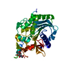
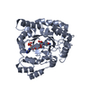
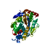
| Entry | Database: PDB / ID: 5ylh | ||||||
|---|---|---|---|---|---|---|---|
| Title | Structure of GH113 beta-1,4-mannanase | ||||||
 Components Components | beta-1,4-mannanase | ||||||
 Keywords Keywords | HYDROLASE / beta-1 / 4-mannanase | ||||||
| Function / homology | : / Glycoside Hydrolase Family 113 / Glycosidases / Glycoside hydrolase superfamily / TIM Barrel / Alpha-Beta Barrel / Alpha Beta / 1,4-beta-xylanase Function and homology information Function and homology information | ||||||
| Biological species |  Amphibacillus xylanus NBRC 15112 (bacteria) Amphibacillus xylanus NBRC 15112 (bacteria) | ||||||
| Method |  X-RAY DIFFRACTION / SYNCHROTRON / MOLECULAR REPLACEMENT / Resolution: 2.29 Å X-RAY DIFFRACTION / SYNCHROTRON / MOLECULAR REPLACEMENT / Resolution: 2.29 Å | ||||||
 Authors Authors | Jiang, Z.Q. / You, X. / Yang, S.Q. / Huang, P. / Ma, J.W. | ||||||
 Citation Citation | Journal: J. Biol. Chem. / Year: 2018 Title: Structural insights into the catalytic mechanism of a novel glycoside hydrolase family 113 beta-1,4-mannanase from Amphibacillus xylanus Authors: You, X. / Qin, Z. / Yan, Q. / Yang, S. / Li, Y. / Jiang, Z. #1: Journal: To Be PublishedTitle: Structure of GH113 beta-1,4-mannanase at 2.3 Angstroms resolution Authors: Jiang, Z.Q. / You, X. / Yang, S.Q. / Huang, P. / Ma, J.W. | ||||||
| History |
|
- Structure visualization
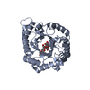
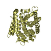
| Structure viewer | Molecule: MolmilJmol/JSmol |
|---|
- Downloads & links
-Download
| PDBx/mmCIF format | 5ylh.cif.gz | 138.2 KB | Display | PDBx/mmCIF format |
|---|---|---|---|---|
| PDB format | pdb5ylh.ent.gz | 108.2 KB | Display | PDB format |
| PDBx/mmJSON format | 5ylh.json.gz | Tree view | PDBx/mmJSON format | |
| Others |  Other downloads Other downloads |
-Validation report
| Arichive directory | https://data.pdbj.org/pub/pdb/validation_reports/yl/5ylhftp://data.pdbj.org/pub/pdb/validation_reports/yl/5ylh | HTTPS FTP |
|---|
-Related structure data
| Related structure data |  5yliC  5ylkC  5yllC  5z4tC 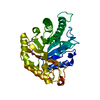 3civS S: Starting model for refinement C: citing same article ( |
|---|---|
| Similar structure data |


-Links
 PDBj
PDBj- Assembly
| Deposited unit | 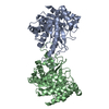
| ||||||||
|---|---|---|---|---|---|---|---|---|---|
| 1 | 
| ||||||||
| 2 | 
| ||||||||
| Unit cell |
|
-Components
| #1: Protein | Mass: 36281.508 Da / Num. of mol.: 2 Source method: isolated from a genetically manipulated source Source: (gene. exp.) Amphibacillus xylanus NBRC 15112 (bacteria)Strain: NBRC 15112 / Gene: AXY_22370 / Production host: #2: Water | ChemComp-HOH / |  Mass: 18.015 Da / Num. of mol.: 160 / Source method: isolated from a natural source / Formula: H2O Mass: 18.015 Da / Num. of mol.: 160 / Source method: isolated from a natural source / Formula: H2O |
|---|
-Experimental details
-Experiment
| Experiment | Method: X-RAY DIFFRACTION / Number of used crystals: 1 |
|---|
- Sample preparation
| Crystal | Density Matthews: 2.48 Å3/Da / Density % sol: 50.31 % |
|---|---|
| Crystal grow | Temperature: 293 K / Method: vapor diffusion, sitting drop / Details: PEG4000, HEPES Na, 2-Propylalcohol |
-Data collection
| Diffraction | Mean temperature: 100 K |
|---|---|
| Diffraction source | Source: SYNCHROTRON / Site: SSRF  / Beamline: BL18U1 / Wavelength: 0.978 Å / Beamline: BL18U1 / Wavelength: 0.978 Å |
| Detector | Type: DECTRIS PILATUS3 S 6M / Detector: PIXEL / Date: Mar 3, 2017 |
| Radiation | Protocol: SINGLE WAVELENGTH / Monochromatic (M) / Laue (L): M / Scattering type: x-ray |
| Radiation wavelength | Wavelength: 0.978 Å / Relative weight: 1 |
| Reflection | Resolution: 2.29→30.38 Å / Num. obs: 29076 / % possible obs: 88 % / Observed criterion σ(I): 1 / Redundancy: 3.7 % / Rmerge(I) obs: 0.12 / Net I/σ(I): 9.8 |
| Reflection shell | Resolution: 2.29→30.383 Å / Redundancy: 3.6 % / Rmerge(I) obs: 0.122 / Mean I/σ(I) obs: 4.6 / Rsym value: 0.118 / % possible all: 88 |
- Processing
| Software |
| ||||||||||||||||
|---|---|---|---|---|---|---|---|---|---|---|---|---|---|---|---|---|---|
| Refinement | Method to determine structure: MOLECULAR REPLACEMENT Starting model: 3CIV Resolution: 2.29→2.38 Å / Cross valid method: FREE R-VALUE
| ||||||||||||||||
| Refinement step | Cycle: LAST / Resolution: 2.29→2.38 Å
| ||||||||||||||||
| LS refinement shell | Highest resolution: 2.29 Å |
