 Movie
Movie Controller

[English] 日本語
 Yorodumi
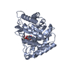
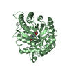
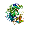
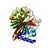
- PDB-3civ: Crystal structure of the endo-beta-1,4-mannanase from Alicyclobac... -
+ Open data

- Basic information
| Entry | Database: PDB / ID: 3civ | ||||||
|---|---|---|---|---|---|---|---|
| Title | Crystal structure of the endo-beta-1,4-mannanase from Alicyclobacillus acidocaldarius | ||||||
 Components Components | Endo-beta-1,4-mannanase | ||||||
 Keywords Keywords | HYDROLASE / TIM BARREL | ||||||
| Function / homology | : / Glycoside Hydrolase Family 113 / Glycosidases / Glycoside hydrolase superfamily / TIM Barrel / Alpha-Beta Barrel / Alpha Beta / Endo-beta-1,4-mannanase Function and homology information Function and homology information | ||||||
| Biological species |  Alicyclobacillus acidocaldarius (bacteria) Alicyclobacillus acidocaldarius (bacteria) | ||||||
| Method |  X-RAY DIFFRACTION / SAD / Resolution: 1.9 Å X-RAY DIFFRACTION / SAD / Resolution: 1.9 Å | ||||||
 Authors Authors | Ma, Y. / Zhang, Y. / Xue, Y. | ||||||
 Citation Citation | Journal: J.Biol.Chem. / Year: 2008 Title: Biochemical and Structural Characterization of the Intracellular Mannanase AaManA of Alicyclobacillus acidocaldarius Reveals a Novel Glycoside Hydrolase Family Belonging to Clan GH-A Authors: Zhang, Y. / Ju, J. / Peng, H. / Gao, F. / Zhou, C. / Zeng, Y. / Xue, Y. / Li, Y. / Henrissat, B. / Gao, G.F. / Ma, Y. | ||||||
| History |
|
- Structure visualization
| Structure viewer | Molecule: MolmilJmol/JSmol |
|---|
- Downloads & links
-Download
| PDBx/mmCIF format | 3civ.cif.gz | 79.5 KB | Display | PDBx/mmCIF format |
|---|---|---|---|---|
| PDB format | pdb3civ.ent.gz | 58.4 KB | Display | PDB format |
| PDBx/mmJSON format | 3civ.json.gz | Tree view | PDBx/mmJSON format | |
| Others |  Other downloads Other downloads |
-Validation report
| Arichive directory | https://data.pdbj.org/pub/pdb/validation_reports/ci/3civftp://data.pdbj.org/pub/pdb/validation_reports/ci/3civ | HTTPS FTP |
|---|
-Related structure data
| Similar structure data |
|---|


-Links
 PDBj
PDBj- Assembly
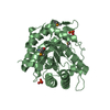
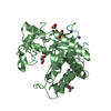
| Deposited unit | 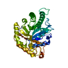
| ||||||||
|---|---|---|---|---|---|---|---|---|---|
| 1 |
| ||||||||
| Unit cell |
|
-Components
| #1: Protein | Mass: 38803.668 Da / Num. of mol.: 1 Source method: isolated from a genetically manipulated source Source: (gene. exp.) Alicyclobacillus acidocaldarius (bacteria)Strain: Tc-12-31 / Gene: AamanA / Plasmid: pET28a / Production host: References: UniProt: A5H1I6, mannan endo-1,4-beta-mannosidase |
|---|---|
| #2: Water | ChemComp-HOH /  Mass: 18.015 Da / Num. of mol.: 221 / Source method: isolated from a natural source / Formula: H2O Mass: 18.015 Da / Num. of mol.: 221 / Source method: isolated from a natural source / Formula: H2O |
-Experimental details
-Experiment
| Experiment | Method: X-RAY DIFFRACTION / Number of used crystals: 1 |
|---|
- Sample preparation
| Crystal | Density Matthews: 1.9 Å3/Da / Density % sol: 35.25 % |
|---|---|
| Crystal grow | Temperature: 291 K / Method: vapor diffusion, hanging drop / pH: 4.6 Details: 0.2M ammonium dihydrogen phosphate, 0.1M sodium citrate (pH 4.6), VAPOR DIFFUSION, HANGING DROP, temperature 291K |
-Data collection
| Diffraction | Mean temperature: 100 K |
|---|---|
| Diffraction source | Source: ROTATING ANODE / Type: RIGAKU MICROMAX-007 / Wavelength: 1.5418 Å |
| Detector | Type: RIGAKU RAXIS VII / Detector: IMAGE PLATE / Details: mirrors |
| Radiation | Protocol: SINGLE WAVELENGTH / Monochromatic (M) / Laue (L): M / Scattering type: x-ray |
| Radiation wavelength | Wavelength: 1.5418 Å / Relative weight: 1 |
| Reflection | Resolution: 1.9→35.07 Å / Num. obs: 23386 / % possible obs: 97.3 % / Redundancy: 6.92 % / Rmerge(I) obs: 0.055 / Net I/σ(I): 20.8 |
| Reflection shell | Resolution: 1.9→1.97 Å / Redundancy: 5.8 % / Rmerge(I) obs: 0.144 / Mean I/σ(I) obs: 10.2 / % possible all: 89.4 |
- Processing
| Software |
| ||||||||||||||||||||||||||||||||||||||||||||||||||||||||||||||||||||||||||||||||||||||||||
|---|---|---|---|---|---|---|---|---|---|---|---|---|---|---|---|---|---|---|---|---|---|---|---|---|---|---|---|---|---|---|---|---|---|---|---|---|---|---|---|---|---|---|---|---|---|---|---|---|---|---|---|---|---|---|---|---|---|---|---|---|---|---|---|---|---|---|---|---|---|---|---|---|---|---|---|---|---|---|---|---|---|---|---|---|---|---|---|---|---|---|---|
| Refinement | Method to determine structure: SAD / Resolution: 1.9→35.07 Å / Cor.coef. Fo:Fc: 0.951 / Cor.coef. Fo:Fc free: 0.934 / SU B: 2.86 / SU ML: 0.087 / Cross valid method: THROUGHOUT / ESU R: 0.168 / ESU R Free: 0.146 / Stereochemistry target values: MAXIMUM LIKELIHOOD / Details: HYDROGENS HAVE BEEN ADDED IN THE RIDING POSITIONS
| ||||||||||||||||||||||||||||||||||||||||||||||||||||||||||||||||||||||||||||||||||||||||||
| Solvent computation | Ion probe radii: 0.8 Å / Shrinkage radii: 0.8 Å / VDW probe radii: 1.4 Å / Solvent model: MASK | ||||||||||||||||||||||||||||||||||||||||||||||||||||||||||||||||||||||||||||||||||||||||||
| Displacement parameters | Biso mean: 16.037 Å2
| ||||||||||||||||||||||||||||||||||||||||||||||||||||||||||||||||||||||||||||||||||||||||||
| Refinement step | Cycle: LAST / Resolution: 1.9→35.07 Å
| ||||||||||||||||||||||||||||||||||||||||||||||||||||||||||||||||||||||||||||||||||||||||||
| Refine LS restraints |
| ||||||||||||||||||||||||||||||||||||||||||||||||||||||||||||||||||||||||||||||||||||||||||
| LS refinement shell | Resolution: 1.9→1.949 Å / Total num. of bins used: 20
|
