 Movie
Movie Controller

[English] 日本語
 Yorodumi
- PDB-1bt1: CATECHOL OXIDASE FROM IPOMOEA BATATAS (SWEET POTATOES) IN THE NAT... -
+ Open data

- Basic information
| Entry | Database: PDB / ID: 1bt1 | ||||||
|---|---|---|---|---|---|---|---|
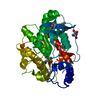
| Title | CATECHOL OXIDASE FROM IPOMOEA BATATAS (SWEET POTATOES) IN THE NATIVE CU(II)-CU(II) STATE | ||||||
 Components Components | PROTEIN (CATECHOL OXIDASE) | ||||||
 Keywords Keywords | OXIDOREDUCTASE / CATECHOL OXIDASE / DICOPPER ENZYME / IPOMOEA BATATAS | ||||||
| Function / homology |  Function and homology information Function and homology informationcatechol oxidase / catechol oxidase activity / chloroplast thylakoid lumen / copper ion binding Similarity search - Function | ||||||
| Biological species |  Ipomoea batatas (sweet potato) Ipomoea batatas (sweet potato) | ||||||
| Method |  X-RAY DIFFRACTION / MIR / Resolution: 2.7 Å X-RAY DIFFRACTION / MIR / Resolution: 2.7 Å | ||||||
 Authors Authors | Klabunde, T. / Eicken, C. / Sacchettini, J.C. / Krebs, B. | ||||||
 Citation Citation | Journal: Nat.Struct.Biol. / Year: 1998 Title: Crystal structure of a plant catechol oxidase containing a dicopper center. Authors: Klabunde, T. / Eicken, C. / Sacchettini, J.C. / Krebs, B. | ||||||
| History |
|
- Structure visualization
| Structure viewer | Molecule: MolmilJmol/JSmol |
|---|
- Downloads & links
-Download
| PDBx/mmCIF format | 1bt1.cif.gz | 144.3 KB | Display | PDBx/mmCIF format |
|---|---|---|---|---|
| PDB format | pdb1bt1.ent.gz | 114.4 KB | Display | PDB format |
| PDBx/mmJSON format | 1bt1.json.gz | Tree view | PDBx/mmJSON format | |
| Others |  Other downloads Other downloads |
-Validation report
| Arichive directory | https://data.pdbj.org/pub/pdb/validation_reports/bt/1bt1ftp://data.pdbj.org/pub/pdb/validation_reports/bt/1bt1 | HTTPS FTP |
|---|
-Related structure data


-Links
 PDBj
PDBj- Assembly
| Deposited unit | 
| ||||||||
|---|---|---|---|---|---|---|---|---|---|
| 1 | 
| ||||||||
| 2 | 
| ||||||||
| Unit cell |
| ||||||||
| Noncrystallographic symmetry (NCS) | NCS oper: (Code: given Matrix: (0.999952, 9.9E-5, 0.00976), Vector: |
-Components
| #1: Protein | Mass: 38814.582 Da / Num. of mol.: 2 / Source method: isolated from a natural source / Details: COVALENT THIOETHER BOND BETWEEN H109 AND C92 / Source: (natural) Ipomoea batatas (sweet potato) / Organ: MATURE TUBER / References: UniProt: Q9ZP19, catechol oxidase#2: Chemical |   Mass: 143.091 Da / Num. of mol.: 2 / Source method: obtained synthetically / Formula: Cu2O Mass: 143.091 Da / Num. of mol.: 2 / Source method: obtained synthetically / Formula: Cu2O#3: Water | ChemComp-HOH / |  Mass: 18.015 Da / Num. of mol.: 168 / Source method: isolated from a natural source / Formula: H2O Mass: 18.015 Da / Num. of mol.: 168 / Source method: isolated from a natural source / Formula: H2OHas protein modification | Y | |
|---|
-Experimental details
-Experiment
| Experiment | Method: X-RAY DIFFRACTION / Number of used crystals: 1 |
|---|
- Sample preparation
| Crystal | Density Matthews: 2.51 Å3/Da / Density % sol: 51.08 % | ||||||||||||||||||||||||||||||||||||
|---|---|---|---|---|---|---|---|---|---|---|---|---|---|---|---|---|---|---|---|---|---|---|---|---|---|---|---|---|---|---|---|---|---|---|---|---|---|
| Crystal grow | Method: vapor diffusion, hanging drop / pH: 7 Details: CRYSTALS WERE GROWN AT 277 K FROM SOLUTIONS CONTAINING 14 MG/ML PROTEIN, 120 MG/ML PEG6000, 500 MM NACL, 50 MM HEPES, PH 7.0, EQUILIBRATED AGAINST A SOLUTION CONTAINING 200 MG/ML PEG6000., ...Details: CRYSTALS WERE GROWN AT 277 K FROM SOLUTIONS CONTAINING 14 MG/ML PROTEIN, 120 MG/ML PEG6000, 500 MM NACL, 50 MM HEPES, PH 7.0, EQUILIBRATED AGAINST A SOLUTION CONTAINING 200 MG/ML PEG6000., VAPOR DIFFUSION, HANGING DROP | ||||||||||||||||||||||||||||||||||||
| Crystal grow | *PLUS Temperature: 4 ℃ | ||||||||||||||||||||||||||||||||||||
| Components of the solutions | *PLUS
|
-Data collection
| Diffraction | Mean temperature: 291 K |
|---|---|
| Diffraction source | Source: ROTATING ANODE / Type: RIGAKU / Wavelength: 1.54 |
| Detector | Type: MACSCIENCE / Detector: IMAGE PLATE / Date: Aug 15, 1997 / Details: MIRRORS |
| Radiation | Monochromator: NI FILTER / Protocol: SINGLE WAVELENGTH / Monochromatic (M) / Laue (L): M / Scattering type: x-ray |
| Radiation wavelength | Wavelength: 1.54 Å / Relative weight: 1 |
| Reflection | Resolution: 2.7→20 Å / Num. obs: 18909 / % possible obs: 90.1 % / Observed criterion σ(I): 0 / Redundancy: 2.4 % / Rsym value: 0.064 / Net I/σ(I): 16.4 |
| Reflection shell | Resolution: 2.7→2.8 Å / Rsym value: 0.127 / % possible all: 92.3 |
| Reflection | *PLUS Num. measured all: 45122 / Rmerge(I) obs: 0.064 |
| Reflection shell | *PLUS % possible obs: 92.3 % / Rmerge(I) obs: 0.127 |
- Processing
| Software |
| ||||||||||||||||||||||||||||||||||||||||||||||||||||||||||||
|---|---|---|---|---|---|---|---|---|---|---|---|---|---|---|---|---|---|---|---|---|---|---|---|---|---|---|---|---|---|---|---|---|---|---|---|---|---|---|---|---|---|---|---|---|---|---|---|---|---|---|---|---|---|---|---|---|---|---|---|---|---|
| Refinement | Method to determine structure: MIR / Resolution: 2.7→8 Å / σ(F): 3 Details: RESIDUES INVOLVED IN PACKING INTERACTIONS WERE EXCLUDED FROM THE NCS RESTRAINTS.
| ||||||||||||||||||||||||||||||||||||||||||||||||||||||||||||
| Displacement parameters | Biso mean: 13 Å2 | ||||||||||||||||||||||||||||||||||||||||||||||||||||||||||||
| Refinement step | Cycle: LAST / Resolution: 2.7→8 Å
| ||||||||||||||||||||||||||||||||||||||||||||||||||||||||||||
| Refine LS restraints |
| ||||||||||||||||||||||||||||||||||||||||||||||||||||||||||||
| Refine LS restraints NCS | NCS model details: RESTRAINTS / Rms dev position: 0.094 Å / Weight position: 100 | ||||||||||||||||||||||||||||||||||||||||||||||||||||||||||||
| Xplor file | Serial no: 1 / Param file: PARHCSDX.PRO / Topol file: TOPHCSDX.PRO | ||||||||||||||||||||||||||||||||||||||||||||||||||||||||||||
| Software | *PLUS Name: X-PLOR / Version: 3.1 / Classification: refinement | ||||||||||||||||||||||||||||||||||||||||||||||||||||||||||||
| Refinement | *PLUS Lowest resolution: 8 Å / σ(F): 3 / % reflection Rfree: 5 % | ||||||||||||||||||||||||||||||||||||||||||||||||||||||||||||
| Solvent computation | *PLUS | ||||||||||||||||||||||||||||||||||||||||||||||||||||||||||||
| Displacement parameters | *PLUS Biso mean: 13 Å2 |
