 Movie
Movie Controller

[English] 日本語
 Yorodumi
- PDB-6ptf: Crystal Structure of CobT from Methanocaldococcus jannaschii in A... -
+ Open data

- Basic information
| Entry | Database: PDB / ID: 6ptf | ||||||
|---|---|---|---|---|---|---|---|
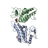
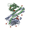
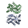
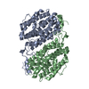
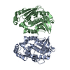
| Title | Crystal Structure of CobT from Methanocaldococcus jannaschii in Apo State | ||||||
 Components Components | UPF0284 protein MJ1598 | ||||||
 Keywords Keywords | TRANSFERASE | ||||||
| Function / homology |  Function and homology information Function and homology informationnicotinate-nucleotide-dimethylbenzimidazole phosphoribosyltransferase activity Similarity search - Function | ||||||
| Biological species |   Methanocaldococcus jannaschii (archaea) Methanocaldococcus jannaschii (archaea) | ||||||
| Method |  X-RAY DIFFRACTION / SYNCHROTRON / MOLECULAR REPLACEMENT / Resolution: 2.203 Å X-RAY DIFFRACTION / SYNCHROTRON / MOLECULAR REPLACEMENT / Resolution: 2.203 Å | ||||||
 Authors Authors | Schwarzwalder, A.H. / Jeter, V.L. / Vecellio, A.A. / Erpenbach, E. / Escalante-Semerena, J.C. / Rayment, I. | ||||||
| Funding support |  United States, 1items United States, 1items
| ||||||
 Citation Citation | Journal: Sci Rep / Year: 2022 Title: Structural studies of the phosphoribosyltransferase involved in cobamide biosynthesis in methanogenic archaea and cyanobacteria. Authors: Jeter, V.L. / Schwarzwalder, A.H. / Rayment, I. / Escalante-Semerena, J.C. | ||||||
| History |
|
- Structure visualization
| Structure viewer | Molecule: MolmilJmol/JSmol |
|---|
- Downloads & links
-Download
| PDBx/mmCIF format | 6ptf.cif.gz | 77.9 KB | Display | PDBx/mmCIF format |
|---|---|---|---|---|
| PDB format | pdb6ptf.ent.gz | 57.4 KB | Display | PDB format |
| PDBx/mmJSON format | 6ptf.json.gz | Tree view | PDBx/mmJSON format | |
| Others |  Other downloads Other downloads |
-Validation report
| Arichive directory | https://data.pdbj.org/pub/pdb/validation_reports/pt/6ptfftp://data.pdbj.org/pub/pdb/validation_reports/pt/6ptf | HTTPS FTP |
|---|
-Related structure data
| Related structure data |  6pt8C  6pu6C  3l0zS S: Starting model for refinement C: citing same article ( |
|---|---|
| Similar structure data |


-Links
 PDBj
PDBj- Assembly
| Deposited unit | 
| ||||||||
|---|---|---|---|---|---|---|---|---|---|
| 1 | 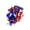
| ||||||||
| Unit cell |
| ||||||||
| Components on special symmetry positions |
|
-Components
| #1: Protein | Mass: 37847.953 Da / Num. of mol.: 1 Source method: isolated from a genetically manipulated source Source: (gene. exp.) Methanocaldococcus jannaschii (strain ATCC 43067 / DSM 2661 / JAL-1 / JCM 10045 / NBRC 100440) (archaea)Strain: ATCC 43067 / DSM 2661 / JAL-1 / JCM 10045 / NBRC 100440 Gene: MJ1598 / Production host:  |
|---|---|
| #2: Water | ChemComp-HOH /  Mass: 18.015 Da / Num. of mol.: 35 / Source method: isolated from a natural source / Formula: H2O Mass: 18.015 Da / Num. of mol.: 35 / Source method: isolated from a natural source / Formula: H2O |
-Experimental details
-Experiment
| Experiment | Method: X-RAY DIFFRACTION / Number of used crystals: 1 |
|---|
- Sample preparation
| Crystal | Density Matthews: 2.53 Å3/Da / Density % sol: 51.42 % |
|---|---|
| Crystal grow | Temperature: 298 K / Method: vapor diffusion, hanging drop / pH: 5.5 Details: Crystals formed by mixing 13.3 mg/ml protein solution with well solution containing 8.4% PEG 8K, 100 mM MES/ acetate buffer pH 5.5, 1 mM 5-hydroxybenzimidazole. Crystals were cryoprotected ...Details: Crystals formed by mixing 13.3 mg/ml protein solution with well solution containing 8.4% PEG 8K, 100 mM MES/ acetate buffer pH 5.5, 1 mM 5-hydroxybenzimidazole. Crystals were cryoprotected by overnight soak in 20% ethylene glycol, 100 mM MES/acetate buffer pH 5.5, 16% Peg 4K and 5 mM 5,6-dimethylbenzimidazole prior to freezing |
-Data collection
| Diffraction | Mean temperature: 100 K / Serial crystal experiment: N |
|---|---|
| Diffraction source | Source: SYNCHROTRON / Site: APS / Beamline: 19-ID / Wavelength: 0.97934 Å |
| Detector | Type: DECTRIS PILATUS3 X 6M / Detector: PIXEL / Date: Aug 15, 2018 |
| Radiation | Protocol: SINGLE WAVELENGTH / Monochromatic (M) / Laue (L): M / Scattering type: x-ray |
| Radiation wavelength | Wavelength: 0.97934 Å / Relative weight: 1 |
| Reflection | Resolution: 2.203→46.1 Å / Num. obs: 20650 / % possible obs: 99.68 % / Redundancy: 12.4 % / CC1/2: 0.999 / Rmerge(I) obs: 0.05481 / Net I/σ(I): 24.59 |
| Reflection shell | Resolution: 2.203→2.282 Å / Redundancy: 12.2 % / Rmerge(I) obs: 0.6916 / Mean I/σ(I) obs: 3.15 / Num. unique obs: 1997 / CC1/2: 0.969 / % possible all: 99.35 |
- Processing
| Software |
| ||||||||||||||||||||||||||||||||||||||||||||||||
|---|---|---|---|---|---|---|---|---|---|---|---|---|---|---|---|---|---|---|---|---|---|---|---|---|---|---|---|---|---|---|---|---|---|---|---|---|---|---|---|---|---|---|---|---|---|---|---|---|---|
| Refinement | Method to determine structure: MOLECULAR REPLACEMENT Starting model: 3L0z Resolution: 2.203→46.097 Å / SU ML: 0.26 / Cross valid method: FREE R-VALUE / σ(F): 1.35 / Phase error: 28.34
| ||||||||||||||||||||||||||||||||||||||||||||||||
| Solvent computation | Shrinkage radii: 0.9 Å / VDW probe radii: 1.11 Å | ||||||||||||||||||||||||||||||||||||||||||||||||
| Displacement parameters | Biso max: 151.86 Å2 / Biso mean: 67.0932 Å2 / Biso min: 34.67 Å2 | ||||||||||||||||||||||||||||||||||||||||||||||||
| Refinement step | Cycle: final / Resolution: 2.203→46.097 Å
| ||||||||||||||||||||||||||||||||||||||||||||||||
| LS refinement shell | Refine-ID: X-RAY DIFFRACTION / Rfactor Rfree error: 0
|
