 Movie
Movie Controller


+ Open data

- Basic information
| Entry | Database: PDB / ID: 6u0v | ||||||
|---|---|---|---|---|---|---|---|
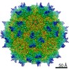
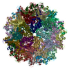
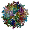
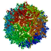
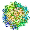
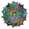
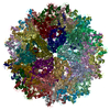
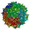
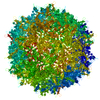
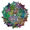
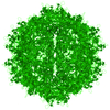
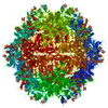
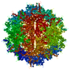
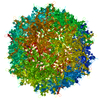
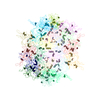
| Title | Atomic-Resolution Cryo-EM Structure of AAV2 VLP | ||||||
 Components Components | Capsid protein VP1 | ||||||
 Keywords Keywords | VIRUS LIKE PARTICLE / Protrusion / axes / receptor / pore | ||||||
| Function / homology |  Function and homology information Function and homology informationsymbiont entry into host cell via permeabilization of host membrane / host cell nucleolus / T=1 icosahedral viral capsid / clathrin-dependent endocytosis of virus by host cell / virion attachment to host cell / structural molecule activity Similarity search - Function | ||||||
| Biological species |  Adeno-associated virus - 2 Adeno-associated virus - 2 | ||||||
| Method | ELECTRON MICROSCOPY / single particle reconstruction / cryo EM / Resolution: 3.02 Å | ||||||
 Authors Authors | Agbandje-Mckenna, M. / Bennett, A. | ||||||
| Funding support |  United States, 1items United States, 1items
| ||||||
 Citation Citation | Journal: J Struct Biol / Year: 2020 Title: Structure comparison of the chimeric AAV2.7m8 vector with parental AAV2. Authors: Antonette Bennett / Annahita Keravala / Victoria Makal / Justin Kurian / Brahim Belbellaa / Rangoli Aeran / Yu-Shan Tseng / Duncan Sousa / John Spear / Mehdi Gasmi / Mavis Agbandje-McKenna / Abstract: The AAV2.7m8 vector is an engineered capsid with a 10-amino acid insertion in adeno-associated virus (AAV) surface variable region VIII (VR-VIII) resulting in the alteration of an antigenic region of ...The AAV2.7m8 vector is an engineered capsid with a 10-amino acid insertion in adeno-associated virus (AAV) surface variable region VIII (VR-VIII) resulting in the alteration of an antigenic region of AAV2 and the ability to efficiently transduce retina cells following intravitreal administration. Directed evolution and in vivo screening in the mouse retina isolated this vector. In the present study, we sought to identify the structural differences between a recombinant AAV2.7m8 (rAAV2.7m8) vector packaging a GFP genome and its parental serotype, AAV2, by cryo-electron microscopy (cryo-EM) and image reconstruction. The structures of rAAV2.7m8 and AAV2 were determined to 2.91 and 3.02 Å resolution, respectively. The rAAV2.7m8 amino acid side-chains for residues 219-745 (the last C-terminal residue) were interpretable in the density map with the exception of the 10 inserted amino acids. While observable in a low sigma threshold density, side-chains were only resolved at the base of the insertion, likely due to flexibility at the top of the loop. A comparison to parental AAV2 (ordered from residues 217-735) showed the structures to be similar, except at some side-chains that had different orientations and, in VR-VIII containing the 10 amino acid insertion. VR-VIII is part of an AAV2 antigenic epitope, and the difference is consistent with rAAV2.7m8's escape from a known AAV2 monoclonal antibody, C37-B. The observations provide valuable insight into the configuration of inserted surface peptides on the AAV capsid and structural differences to be leveraged for future AAV vector rational design, especially for retargeted tropism and antibody escape. | ||||||
| History |
|
- Structure visualization
| Movie |
Movie viewer |
|---|---|
| Structure viewer | Molecule: MolmilJmol/JSmol |
- Downloads & links
-Download
| PDBx/mmCIF format | 6u0v.cif.gz | 5.7 MB | Display | PDBx/mmCIF format |
|---|---|---|---|---|
| PDB format | pdb6u0v.ent.gz | Display | PDB format | |
| PDBx/mmJSON format | 6u0v.json.gz | Tree view | PDBx/mmJSON format | |
| Others |  Other downloads Other downloads |
-Validation report
| Arichive directory | https://data.pdbj.org/pub/pdb/validation_reports/u0/6u0vftp://data.pdbj.org/pub/pdb/validation_reports/u0/6u0v | HTTPS FTP |
|---|
-Related structure data
| Related structure data | 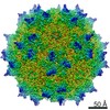 20610MC  6u0rC M: map data used to model this data C: citing same article ( |
|---|---|
| Similar structure data |


-Links
 PDBj
PDBj


- Assembly
| Deposited unit | 
|
|---|---|
| 1 |
|
-Components
| #1: Protein | Mass: 59129.949 Da / Num. of mol.: 60 Source method: isolated from a genetically manipulated source Source: (gene. exp.) Adeno-associated virus - 2 / Gene: VP1 / Cell line (production host): SF9 / Production host:   Spodoptera frugiperda (fall armyworm) / References: UniProt: P03135 Spodoptera frugiperda (fall armyworm) / References: UniProt: P03135 |
|---|
-Experimental details
-Experiment
| Experiment | Method: ELECTRON MICROSCOPY |
|---|---|
| EM experiment | Aggregation state: PARTICLE / 3D reconstruction method: single particle reconstruction |
- Sample preparation
| Component | Name: Adeno-associated virus - 2 / Type: VIRUS / Entity ID: all / Source: RECOMBINANT |
|---|---|
| Molecular weight | Value: 4 MDa / Experimental value: NO |
| Source (natural) | Organism: Adeno-associated virus - 2 |
| Source (recombinant) | Organism: Spodoptera frugiperda (fall armyworm) / Cell: SF9 |
| Details of virus | Empty: YES / Enveloped: NO / Isolate: SEROTYPE / Type: VIRUS-LIKE PARTICLE |
| Virus shell | Diameter: 260 nm / Triangulation number (T number): 1 |
| Buffer solution | pH: 7.4 |
| Specimen | Conc.: 1 mg/ml / Embedding applied: NO / Shadowing applied: NO / Staining applied: NO / Vitrification applied: YES |
| Specimen support | Details: unspecified |
| Vitrification | Instrument: FEI VITROBOT MARK IV / Cryogen name: ETHANE |
- Electron microscopy imaging
| Experimental equipment |  Model: Titan Krios / Image courtesy: FEI Company |
|---|---|
| Microscopy | Model: FEI TITAN KRIOS |
| Electron gun | Electron source:  FIELD EMISSION GUN / Accelerating voltage: 300 kV / Illumination mode: FLOOD BEAM FIELD EMISSION GUN / Accelerating voltage: 300 kV / Illumination mode: FLOOD BEAM |
| Electron lens | Mode: BRIGHT FIELD |
| Image recording | Electron dose: 75 e/Å2 / Detector mode: COUNTING / Film or detector model: GATAN K2 SUMMIT (4k x 4k) / Num. of real images: 410 |
- Processing
| EM software |
| |||||||||
|---|---|---|---|---|---|---|---|---|---|---|
| CTF correction | Type: PHASE FLIPPING AND AMPLITUDE CORRECTION | |||||||||
| Particle selection | Num. of particles selected: 13557 | |||||||||
| 3D reconstruction | Resolution: 3.02 Å / Resolution method: FSC 0.143 CUT-OFF / Num. of particles: 6778 / Symmetry type: POINT | |||||||||
| Atomic model building | B value: 100 / Protocol: FLEXIBLE FIT |
