 Movie
Movie Controller

[English] 日本語
 Yorodumi
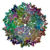
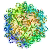
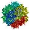
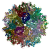
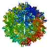
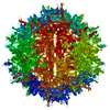
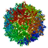
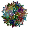
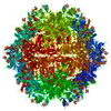
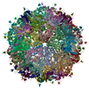
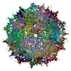
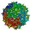
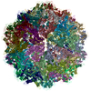
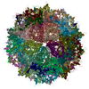
- PDB-6u95: Adeno-associated virus strain AAVhu.37 capsid icosahedral structure -
+ Open data

- Basic information
| Entry | Database: PDB / ID: 6u95 | ||||||
|---|---|---|---|---|---|---|---|
| Title | Adeno-associated virus strain AAVhu.37 capsid icosahedral structure | ||||||
 Components Components | Capsid protein VP1 | ||||||
 Keywords Keywords | VIRUS / capsid / blood-brain barrier penetration | ||||||
| Function / homology |  Function and homology information Function and homology information | ||||||
| Biological species |   Adeno-associated virus Adeno-associated virus | ||||||
| Method | ELECTRON MICROSCOPY / single particle reconstruction / cryo EM / Resolution: 2.56 Å | ||||||
 Authors Authors | Kaelber, J.T. / Yost, S.A. / Firlar, E. / Mercer, A.C. | ||||||
 Citation Citation | Journal: Acta Crystallogr F Struct Biol Commun / Year: 2020 Title: Structure of the AAVhu.37 capsid by cryoelectron microscopy. Authors: Jason T Kaelber / Samantha A Yost / Keith A Webber / Emre Firlar / Ye Liu / Olivier Danos / Andrew C Mercer /  Abstract: Adeno-associated viruses (AAVs) are used as in vivo gene-delivery vectors in gene-therapy products and have been heavily investigated for numerous indications. Over 100 naturally occurring AAV ...Adeno-associated viruses (AAVs) are used as in vivo gene-delivery vectors in gene-therapy products and have been heavily investigated for numerous indications. Over 100 naturally occurring AAV serotypes and variants have been isolated from primate samples. Many reports have described unique properties of these variants (for instance, differences in potency, target cell or evasion of the immune response), despite high amino-acid sequence conservation. AAVhu.37 is of interest for clinical applications owing to its proficient transduction of the liver and central nervous system. The sequence identity of the AAVhu.37 VP1 to the well characterized AAVrh.10 serotype, for which no structure is available, is greater than 98%. Here, the structure of the AAVhu.37 capsid at 2.56 Å resolution obtained via single-particle cryo-electron microscopy is presented. | ||||||
| History |
|
- Structure visualization
| Movie |
Movie viewer |
|---|---|
| Structure viewer | Molecule: MolmilJmol/JSmol |
- Downloads & links
-Download
| PDBx/mmCIF format | 6u95.cif.gz | 120.1 KB | Display | PDBx/mmCIF format |
|---|---|---|---|---|
| PDB format | pdb6u95.ent.gz | 87.1 KB | Display | PDB format |
| PDBx/mmJSON format | 6u95.json.gz | Tree view | PDBx/mmJSON format | |
| Others |  Other downloads Other downloads |
-Validation report
| Arichive directory | https://data.pdbj.org/pub/pdb/validation_reports/u9/6u95ftp://data.pdbj.org/pub/pdb/validation_reports/u9/6u95 | HTTPS FTP |
|---|
-Related structure data
| Related structure data | 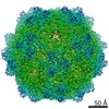 20693MC  7jotC M: map data used to model this data C: citing same article ( |
|---|---|
| Similar structure data |


-Links
 PDBj
PDBj


- Assembly
| Deposited unit | 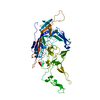
|
|---|---|
| 1 | x 60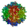
|
| 2 |
|
| 3 | x 5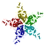
|
| 4 | x 6
|
| 5 | 
|
| Symmetry | Point symmetry: (Schoenflies symbol: I (icosahedral)) |
-Components
| #1: Protein | Mass: 81528.852 Da / Num. of mol.: 1 Source method: isolated from a genetically manipulated source Source: (gene. exp.) Adeno-associated virus / Gene: cap / Cell line (production host): HEK293T / Production host:  Homo sapiens (human) / References: UniProt: Q6JC19 Homo sapiens (human) / References: UniProt: Q6JC19 |
|---|
-Experimental details
-Experiment
| Experiment | Method: ELECTRON MICROSCOPY |
|---|---|
| EM experiment | Aggregation state: PARTICLE / 3D reconstruction method: single particle reconstruction |
- Sample preparation
| Component | Name: Adeno-associated virus / Type: VIRUS Details: Virions containing GFP-encoding ssDNA were assembled in transfected HEK293T helper cells. Entity ID: all / Source: RECOMBINANT | ||||||||||||||||||||
|---|---|---|---|---|---|---|---|---|---|---|---|---|---|---|---|---|---|---|---|---|---|
| Molecular weight | Value: 5 MDa / Experimental value: NO | ||||||||||||||||||||
| Source (natural) | Organism: Adeno-associated virus / Strain: hu.37 | ||||||||||||||||||||
| Source (recombinant) | Organism: Homo sapiens (human) / Strain: HEK293T / Cell: embryonic kidney | ||||||||||||||||||||
| Details of virus | Empty: NO / Enveloped: NO / Isolate: STRAIN / Type: VIRION | ||||||||||||||||||||
| Natural host | Organism: Homo sapiens | ||||||||||||||||||||
| Virus shell | Diameter: 250 nm / Triangulation number (T number): 1 | ||||||||||||||||||||
| Buffer solution | pH: 7.4 / Details: PBS | ||||||||||||||||||||
| Buffer component |
| ||||||||||||||||||||
| Specimen | Embedding applied: NO / Shadowing applied: NO / Staining applied: NO / Vitrification applied: YES / Details: approx. 7*10^13 genome copies per mL | ||||||||||||||||||||
| Specimen support | Grid material: COPPER / Grid mesh size: 200 divisions/in. / Grid type: Quantifoil R2/1 | ||||||||||||||||||||
| Vitrification | Instrument: LEICA EM GP / Cryogen name: ETHANE / Humidity: 100 % / Chamber temperature: 298 K Details: Whatman #1 filter paper, 4.5s blot. 3.5 microliters specimen applied. |
- Electron microscopy imaging
| Experimental equipment |  Model: Talos Arctica / Image courtesy: FEI Company |
|---|---|
| Microscopy | Model: FEI TALOS ARCTICA |
| Electron gun | Electron source:  FIELD EMISSION GUN / Accelerating voltage: 200 kV / Illumination mode: FLOOD BEAM FIELD EMISSION GUN / Accelerating voltage: 200 kV / Illumination mode: FLOOD BEAM |
| Electron lens | Mode: BRIGHT FIELD / Nominal magnification: 130000 X / Calibrated defocus min: 535 nm / Calibrated defocus max: 1584 nm / Cs: 2.7 mm / C2 aperture diameter: 50 µm / Alignment procedure: ZEMLIN TABLEAU |
| Specimen holder | Cryogen: NITROGEN / Specimen holder model: FEI TITAN KRIOS AUTOGRID HOLDER |
| Image recording | Average exposure time: 5 sec. / Electron dose: 1.31 e/Å2 / Detector mode: COUNTING / Film or detector model: GATAN K2 SUMMIT (4k x 4k) / Num. of grids imaged: 1 / Details: collected in superresolution mode |
| EM imaging optics | Energyfilter name: GIF Bioquantum / Chromatic aberration corrector: none / Energyfilter slit width: 20 eV / Spherical aberration corrector: none |
| Image scans | Width: 3838 / Height: 3710 / Movie frames/image: 25 / Used frames/image: 1-25 |
- Processing
| Software | Name: PHENIX / Version: dev_3366: / Classification: refinement | ||||||||||||||||||||||||||||||||||||
|---|---|---|---|---|---|---|---|---|---|---|---|---|---|---|---|---|---|---|---|---|---|---|---|---|---|---|---|---|---|---|---|---|---|---|---|---|---|
| EM software |
| ||||||||||||||||||||||||||||||||||||
| CTF correction | Type: PHASE FLIPPING AND AMPLITUDE CORRECTION | ||||||||||||||||||||||||||||||||||||
| Particle selection | Num. of particles selected: 53545 | ||||||||||||||||||||||||||||||||||||
| Symmetry | Point symmetry: I (icosahedral) | ||||||||||||||||||||||||||||||||||||
| 3D reconstruction | Resolution: 2.56 Å / Resolution method: FSC 0.143 CUT-OFF / Num. of particles: 41850 / Algorithm: FOURIER SPACE Details: 2.8 unmasked, 2.53 masked, 2.56 with noise-substitution ("true FSC"). 2.7 3-bit unmasked. Symmetry type: POINT | ||||||||||||||||||||||||||||||||||||
| Atomic model building | Protocol: FLEXIBLE FIT | ||||||||||||||||||||||||||||||||||||
| Atomic model building | PDB-ID: 2QA0 Pdb chain-ID: A / Accession code: 2QA0 / Pdb chain residue range: 220-738 / Source name: PDB / Type: experimental model | ||||||||||||||||||||||||||||||||||||
| Refine LS restraints |
|

