 Movie
Movie Controller


+ Open data

- Basic information
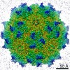
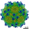
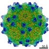
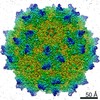
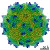
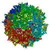
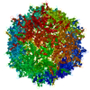
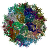
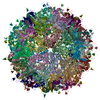
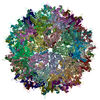
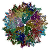
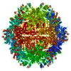
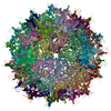
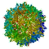
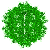
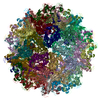
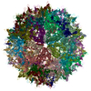
| Entry | Database: PDB / ID: 7l6a | ||||||||||||||||||||||||||||||
|---|---|---|---|---|---|---|---|---|---|---|---|---|---|---|---|---|---|---|---|---|---|---|---|---|---|---|---|---|---|---|---|
| Title | The genome-containing AAV12 capsid | ||||||||||||||||||||||||||||||
 Components Components | VP1 | ||||||||||||||||||||||||||||||
 Keywords Keywords | VIRUS / Icosahedral Capsid / AAV12 / Adeno-associated virus / Parvovirus / Gene Therapy | ||||||||||||||||||||||||||||||
| Function / homology |  Function and homology information Function and homology information | ||||||||||||||||||||||||||||||
| Biological species |  Adeno-associated virus 12 Adeno-associated virus 12 | ||||||||||||||||||||||||||||||
| Method | ELECTRON MICROSCOPY / single particle reconstruction / cryo EM / Resolution: 2.67 Å | ||||||||||||||||||||||||||||||
 Authors Authors | Mietzsch, M. / Agbandje-McKenna, M. | ||||||||||||||||||||||||||||||
| Funding support |  United States, 1items United States, 1items
| ||||||||||||||||||||||||||||||
 Citation Citation | Journal: Viruses / Year: 2021 Title: Completion of the AAV Structural Atlas: Serotype Capsid Structures Reveals Clade-Specific Features. Authors: Mario Mietzsch / Ariana Jose / Paul Chipman / Nilakshee Bhattacharya / Nadia Daneshparvar / Robert McKenna / Mavis Agbandje-McKenna / Abstract: The capsid structures of most Adeno-associated virus (AAV) serotypes, already assigned to an antigenic clade, have been previously determined. This study reports the remaining capsid structures of ...The capsid structures of most Adeno-associated virus (AAV) serotypes, already assigned to an antigenic clade, have been previously determined. This study reports the remaining capsid structures of AAV7, AAV11, AAV12, and AAV13 determined by cryo-electron microscopy and three-dimensional image reconstruction to 2.96, 2.86, 2.54, and 2.76 Å resolution, respectively. These structures complete the structural atlas of the AAV serotype capsids. AAV7 represents the first clade D capsid structure; AAV11 and AAV12 are of a currently unassigned clade that would include AAV4; and AAV13 represents the first AAV2-AAV3 hybrid clade C capsid structure. These newly determined capsid structures all exhibit the AAV capsid features including 5-fold channels, 3-fold protrusions, 2-fold depressions, and a nucleotide binding pocket with an ordered nucleotide in genome-containing capsids. However, these structures have viral proteins that display clade-specific loop conformations. This structural characterization completes our three-dimensional library of the current AAV serotypes to provide an atlas of surface loop configurations compatible with capsid assembly and amenable for future vector engineering efforts. Derived vectors could improve gene delivery success with respect to specific tissue targeting, transduction efficiency, antigenicity or receptor retargeting. | ||||||||||||||||||||||||||||||
| History |
|
- Structure visualization
| Movie |
Movie viewer |
|---|---|
| Structure viewer | Molecule: MolmilJmol/JSmol |
- Downloads & links
-Download
| PDBx/mmCIF format | 7l6a.cif.gz | 5.2 MB | Display | PDBx/mmCIF format |
|---|---|---|---|---|
| PDB format | pdb7l6a.ent.gz | Display | PDB format | |
| PDBx/mmJSON format | 7l6a.json.gz | Tree view | PDBx/mmJSON format | |
| Others |  Other downloads Other downloads |
-Validation report
| Arichive directory | https://data.pdbj.org/pub/pdb/validation_reports/l6/7l6aftp://data.pdbj.org/pub/pdb/validation_reports/l6/7l6a | HTTPS FTP |
|---|
-Related structure data
| Related structure data | 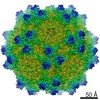 23200MC  7l5qC  7l5uC  7l6bC  7l6eC  7l6fC  7l6hC  7l6iC M: map data used to model this data C: citing same article ( |
|---|---|
| Similar structure data |


-Links
 PDBj
PDBj


- Assembly
| Deposited unit | 
|
|---|---|
| 1 |
|
-Components
| #1: Protein | Mass: 58549.543 Da / Num. of mol.: 60 Source method: isolated from a genetically manipulated source Source: (gene. exp.) Adeno-associated virus 12 / Cell line (production host): HEK293 / Production host:  Homo sapiens (human) / References: UniProt: A9RAI0 Homo sapiens (human) / References: UniProt: A9RAI0#2: Chemical | ChemComp-D5M / 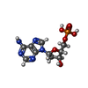  Mass: 331.222 Da / Num. of mol.: 60 / Source method: obtained synthetically / Formula: C10H14N5O6P Mass: 331.222 Da / Num. of mol.: 60 / Source method: obtained synthetically / Formula: C10H14N5O6PHas ligand of interest | N | Has protein modification | N | |
|---|
-Experimental details
-Experiment
| Experiment | Method: ELECTRON MICROSCOPY |
|---|---|
| EM experiment | Aggregation state: PARTICLE / 3D reconstruction method: single particle reconstruction |
- Sample preparation
| Component | Name: Adeno-associated virus 12 / Type: VIRUS / Entity ID: #1 / Source: RECOMBINANT |
|---|---|
| Source (natural) | Organism: Adeno-associated virus 12 |
| Source (recombinant) | Organism: Homo sapiens (human) / Cell: HEK293 |
| Details of virus | Empty: NO / Enveloped: NO / Isolate: OTHER / Type: VIRION |
| Buffer solution | pH: 7.4 |
| Specimen | Embedding applied: NO / Shadowing applied: NO / Staining applied: NO / Vitrification applied: YES |
| Vitrification | Cryogen name: ETHANE |
- Electron microscopy imaging
| Experimental equipment |  Model: Titan Krios / Image courtesy: FEI Company |
|---|---|
| Microscopy | Model: FEI TITAN KRIOS |
| Electron gun | Electron source:  FIELD EMISSION GUN / Accelerating voltage: 300 kV / Illumination mode: FLOOD BEAM FIELD EMISSION GUN / Accelerating voltage: 300 kV / Illumination mode: FLOOD BEAM |
| Electron lens | Mode: BRIGHT FIELD / Cs: 2.7 mm |
| Image recording | Electron dose: 60 e/Å2 / Film or detector model: GATAN K3 (6k x 4k) |
- Processing
| Software | Name: PHENIX / Version: 1.10-2155_2155: / Classification: refinement | ||||||||||||||||||||||||
|---|---|---|---|---|---|---|---|---|---|---|---|---|---|---|---|---|---|---|---|---|---|---|---|---|---|
| EM software |
| ||||||||||||||||||||||||
| CTF correction | Type: PHASE FLIPPING AND AMPLITUDE CORRECTION | ||||||||||||||||||||||||
| 3D reconstruction | Resolution: 2.67 Å / Resolution method: FSC 0.143 CUT-OFF / Num. of particles: 40764 / Symmetry type: POINT | ||||||||||||||||||||||||
| Refine LS restraints |
|
