 Movie
Movie Controller

[English] 日本語
 Yorodumi
- PDB-3ng9: Structure to Function Correlations for Adeno-associated Virus Ser... -
+ Open data

- Basic information
| Entry | Database: PDB / ID: 3ng9 | ||||||
|---|---|---|---|---|---|---|---|
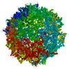
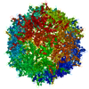
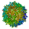
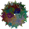
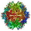
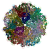
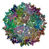
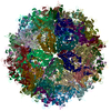
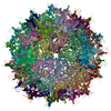
| Title | Structure to Function Correlations for Adeno-associated Virus Serotype 1 | ||||||
 Components Components | Capsid protein | ||||||
 Keywords Keywords | VIRUS / Adeno-Associated Virus 1 / beta barrel / single-stranded DNA virus / parvovirus / icosahedral virus | ||||||
| Function / homology |  Function and homology information Function and homology information | ||||||
| Biological species |  Adeno-associated virus - 1 Adeno-associated virus - 1 | ||||||
| Method |  X-RAY DIFFRACTION / SYNCHROTRON / MOLECULAR REPLACEMENT / Resolution: 2.5 Å X-RAY DIFFRACTION / SYNCHROTRON / MOLECULAR REPLACEMENT / Resolution: 2.5 Å | ||||||
 Authors Authors | Govindasamy, L. / Miller, E.B. / Gurda, B. / McKenna, R. / Zolotukhin, S. / Muzyczka, N. / Agbandje-McKenna, M. | ||||||
 Citation Citation | Journal: To be Published Title: Structure to Function Correlations for Adeno-Associated Virus serotype 1 Authors: Govindasamy, L. / Miller, E.B. / Gurda, B. / McKenna, R. / Zolotukhin, S. / Muzyczka, N. / Agbandje-McKenna, M. | ||||||
| History |
|
- Structure visualization
| Structure viewer | Molecule: MolmilJmol/JSmol |
|---|
- Downloads & links
-Download
| PDBx/mmCIF format | 3ng9.cif.gz | 1 MB | Display | PDBx/mmCIF format |
|---|---|---|---|---|
| PDB format | pdb3ng9.ent.gz | 856.2 KB | Display | PDB format |
| PDBx/mmJSON format | 3ng9.json.gz | Tree view | PDBx/mmJSON format | |
| Others |  Other downloads Other downloads |
-Validation report
| Arichive directory | https://data.pdbj.org/pub/pdb/validation_reports/ng/3ng9ftp://data.pdbj.org/pub/pdb/validation_reports/ng/3ng9 | HTTPS FTP |
|---|
-Related structure data
| Related structure data | 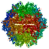 2g8gS S: Starting model for refinement |
|---|---|
| Similar structure data |


-Links
 PDBj
PDBj


- Assembly
| Deposited unit | 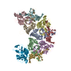
| |||||||||||||||||||||||||||||||||
|---|---|---|---|---|---|---|---|---|---|---|---|---|---|---|---|---|---|---|---|---|---|---|---|---|---|---|---|---|---|---|---|---|---|---|
| 1 | x 6
| |||||||||||||||||||||||||||||||||
| 2 |
| |||||||||||||||||||||||||||||||||
| 3 | 
| |||||||||||||||||||||||||||||||||
| 4 | 
| |||||||||||||||||||||||||||||||||
| Unit cell |
| |||||||||||||||||||||||||||||||||
| Symmetry | Point symmetry: (Schoenflies symbol: I (icosahedral)) | |||||||||||||||||||||||||||||||||
| Components on special symmetry positions |
| |||||||||||||||||||||||||||||||||
| Noncrystallographic symmetry (NCS) | NCS domain:
|
-Components
| #1: Protein | Mass: 81454.922 Da / Num. of mol.: 10 Source method: isolated from a genetically manipulated source Source: (gene. exp.) Adeno-associated virus - 1 / References: UniProt: Q9WBP8#2: Chemical | ChemComp-ADE /   Mass: 135.127 Da / Num. of mol.: 10 / Source method: obtained synthetically / Formula: C5H5N5 Mass: 135.127 Da / Num. of mol.: 10 / Source method: obtained synthetically / Formula: C5H5N5#3: Chemical | ChemComp-CYT /   Mass: 111.102 Da / Num. of mol.: 10 / Source method: obtained synthetically / Formula: C4H5N3O Mass: 111.102 Da / Num. of mol.: 10 / Source method: obtained synthetically / Formula: C4H5N3O#4: Water | ChemComp-HOH / |  Mass: 18.015 Da / Num. of mol.: 1300 / Source method: isolated from a natural source / Formula: H2O Mass: 18.015 Da / Num. of mol.: 1300 / Source method: isolated from a natural source / Formula: H2O |
|---|
-Experimental details
-Experiment
| Experiment | Method: X-RAY DIFFRACTION / Number of used crystals: 6 |
|---|
- Sample preparation
| Crystal | Density Matthews: 2.5 Å3/Da / Density % sol: 50.72 % |
|---|---|
| Crystal grow | Temperature: 298 K / Method: vapor diffusion, hanging drop / pH: 7.3 Details: 1.0 M NaCl and 7% PEG 6K, pH 7.3, VAPOR DIFFUSION, HANGING DROP, temperature 298K |
-Data collection
| Diffraction | Mean temperature: 100 K |
|---|---|
| Diffraction source | Source: SYNCHROTRON / Site: APS  / Beamline: 22-ID / Wavelength: 1 Å / Beamline: 22-ID / Wavelength: 1 Å |
| Detector | Type: MARMOSAIC 300 mm CCD / Detector: CCD |
| Radiation | Monochromator: graphite / Protocol: SINGLE WAVELENGTH / Monochromatic (M) / Laue (L): M / Scattering type: x-ray |
| Radiation wavelength | Wavelength: 1 Å / Relative weight: 1 |
| Reflection | Resolution: 2.5→50 Å / Num. obs: 247785 / % possible obs: 89 % / Redundancy: 3.9 % / Rmerge(I) obs: 0.129 / Rsym value: 0.123 / Net I/σ(I): 12.3 |
| Reflection shell | Resolution: 2.5→2.59 Å / Redundancy: 1.5 % / Rmerge(I) obs: 0.264 / Mean I/σ(I) obs: 3.3 / Num. unique all: 13401 / Rsym value: 0.206 / % possible all: 48.6 |
- Processing
| Software |
| ||||||||||||||||||||||||||||||||||||||||||||||||||||||||||||||||||||||||||||||||||||
|---|---|---|---|---|---|---|---|---|---|---|---|---|---|---|---|---|---|---|---|---|---|---|---|---|---|---|---|---|---|---|---|---|---|---|---|---|---|---|---|---|---|---|---|---|---|---|---|---|---|---|---|---|---|---|---|---|---|---|---|---|---|---|---|---|---|---|---|---|---|---|---|---|---|---|---|---|---|---|---|---|---|---|---|---|---|
| Refinement | Method to determine structure: MOLECULAR REPLACEMENT Starting model: 2G8G Resolution: 2.5→50 Å / Cor.coef. Fo:Fc: 0.91 / Cor.coef. Fo:Fc free: 0.89 / SU B: 9.324 / SU ML: 0.206 / Cross valid method: THROUGHOUT / ESU R Free: 0.284 / Stereochemistry target values: MAXIMUM LIKELIHOOD
| ||||||||||||||||||||||||||||||||||||||||||||||||||||||||||||||||||||||||||||||||||||
| Solvent computation | Ion probe radii: 0.8 Å / Shrinkage radii: 0.8 Å / VDW probe radii: 1.4 Å / Solvent model: BABINET MODEL WITH MASK | ||||||||||||||||||||||||||||||||||||||||||||||||||||||||||||||||||||||||||||||||||||
| Displacement parameters | Biso mean: 36.488 Å2
| ||||||||||||||||||||||||||||||||||||||||||||||||||||||||||||||||||||||||||||||||||||
| Refinement step | Cycle: LAST / Resolution: 2.5→50 Å
| ||||||||||||||||||||||||||||||||||||||||||||||||||||||||||||||||||||||||||||||||||||
| Refine LS restraints |
| ||||||||||||||||||||||||||||||||||||||||||||||||||||||||||||||||||||||||||||||||||||
| Refine LS restraints NCS | Dom-ID: 1 / Ens-ID: 1 / Number: 4260 / Refine-ID: X-RAY DIFFRACTION
| ||||||||||||||||||||||||||||||||||||||||||||||||||||||||||||||||||||||||||||||||||||
| LS refinement shell | Resolution: 2.5→2.565 Å / Total num. of bins used: 20
|
