 Movie
Movie Controller

[English] 日本語
 Yorodumi
- PDB-4v86: Structure-function Analysis of Receptor-binding in Adeno-Associat... -
+ Open data

- Basic information
| Entry | Database: PDB / ID: 4v86 | |||||||||
|---|---|---|---|---|---|---|---|---|---|---|
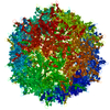
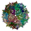
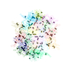
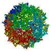
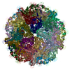
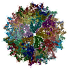
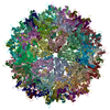
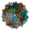
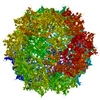
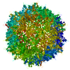
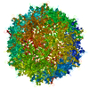
| Title | Structure-function Analysis of Receptor-binding in Adeno-Associated Virus Serotype 6 (AAV-6) | |||||||||
 Components Components | Capsid protein VP1 | |||||||||
 Keywords Keywords | VIRUS / beta barrel | |||||||||
| Function / homology | Phospholipase A2-like domain / Phospholipase A2-like domain / Parvovirus coat protein VP2 / Parvovirus coat protein VP1/VP2 / Parvovirus coat protein VP1/VP2 / Capsid/spike protein, ssDNA virus / T=1 icosahedral viral capsid / structural molecule activity / Capsid protein VP1 Function and homology information Function and homology information | |||||||||
| Biological species |  Adeno-associated virus - 6 Adeno-associated virus - 6 | |||||||||
| Method |  X-RAY DIFFRACTION / SYNCHROTRON / MOLECULAR REPLACEMENT / molecular replacement / Resolution: 3.003 Å X-RAY DIFFRACTION / SYNCHROTRON / MOLECULAR REPLACEMENT / molecular replacement / Resolution: 3.003 Å | |||||||||
 Authors Authors | Xie, Q. | |||||||||
 Citation Citation | Journal: Virology / Year: 2011 Title: Structure-function analysis of receptor-binding in adeno-associated virus serotype 6 (AAV-6). Authors: Xie, Q. / Lerch, T.F. / Meyer, N.L. / Chapman, M.S. | |||||||||
| History |
|
- Structure visualization
| Structure viewer | Molecule: MolmilJmol/JSmol |
|---|
- Downloads & links
-Download
| PDBx/mmCIF format | 4v86.cif.gz | 5.6 MB | Display | PDBx/mmCIF format |
|---|---|---|---|---|
| PDB format | pdb4v86.ent.gz | Display | PDB format | |
| PDBx/mmJSON format | 4v86.json.gz | Tree view | PDBx/mmJSON format | |
| Others |  Other downloads Other downloads |
-Validation report
| Arichive directory | https://data.pdbj.org/pub/pdb/validation_reports/v8/4v86ftp://data.pdbj.org/pub/pdb/validation_reports/v8/4v86 | HTTPS FTP |
|---|
-Related structure data


-Links
 PDBj
PDBj


- Assembly
| Deposited unit | 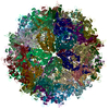
| ||||||||
|---|---|---|---|---|---|---|---|---|---|
| 1 |
| ||||||||
| Unit cell |
|
-Components
| #1: Protein | Mass: 58360.598 Da / Num. of mol.: 60 / Fragment: UNP residues 217-736 Source method: isolated from a genetically manipulated source Source: (gene. exp.) Adeno-associated virus - 6 / Cell line (production host): HELA / Production host:  Homo sapiens (human) / References: UniProt: O56137 Homo sapiens (human) / References: UniProt: O56137 |
|---|
-Experimental details
-Experiment
| Experiment | Method: X-RAY DIFFRACTION / Number of used crystals: 1 |
|---|
- Sample preparation
| Crystal | Density Matthews: 3.43 Å3/Da / Density % sol: 64.12 % |
|---|---|
| Crystal grow | Temperature: 298 K / Method: vapor diffusion, hanging drop / pH: 7.3 Details: 4-6% PEG6000, 100 mM HEPES, 50 mM magnesium chloride,, pH 7.3, VAPOR DIFFUSION, HANGING DROP, temperature 298K |
-Data collection
| Diffraction | Mean temperature: 100 K |
|---|---|
| Diffraction source | Source: SYNCHROTRON / Site: CHESS  / Beamline: F1 / Wavelength: 0.9186 Å / Beamline: F1 / Wavelength: 0.9186 Å |
| Detector | Type: ADSC QUANTUM 270 / Detector: CCD / Date: Aug 1, 2004 |
| Radiation | Monochromator: Single-crystal Si(111) / Protocol: SINGLE WAVELENGTH / Monochromatic (M) / Laue (L): M / Scattering type: x-ray |
| Radiation wavelength | Wavelength: 0.9186 Å / Relative weight: 1 |
| Reflection | Resolution: 3.003→49.21 Å / Num. all: 332221 / Num. obs: 332221 / % possible obs: 42 % / Rmerge(I) obs: 0.12 / Rsym value: 0.12 / Net I/σ(I): 3.7 |
-Phasing
| Phasing | Method: molecular replacement |
|---|
- Processing
| Software |
| |||||||||||||||||||||||||||||||||||
|---|---|---|---|---|---|---|---|---|---|---|---|---|---|---|---|---|---|---|---|---|---|---|---|---|---|---|---|---|---|---|---|---|---|---|---|---|
| Refinement | Method to determine structure: MOLECULAR REPLACEMENT / Resolution: 3.003→49.21 Å / Occupancy max: 1 / Occupancy min: 1 / SU ML: 0.96 / σ(F): 0 / Phase error: 31.46 / Stereochemistry target values: ML
| |||||||||||||||||||||||||||||||||||
| Solvent computation | Shrinkage radii: 0.27 Å / VDW probe radii: 0.6 Å / Solvent model: FLAT BULK SOLVENT MODEL / Bsol: 28.443 Å2 / ksol: 0.328 e/Å3 | |||||||||||||||||||||||||||||||||||
| Displacement parameters | Biso max: 149.19 Å2 / Biso mean: 62.7664 Å2 / Biso min: 35.63 Å2
| |||||||||||||||||||||||||||||||||||
| Refinement step | Cycle: LAST / Resolution: 3.003→49.21 Å
| |||||||||||||||||||||||||||||||||||
| Refine LS restraints |
| |||||||||||||||||||||||||||||||||||
| LS refinement shell | Refine-ID: X-RAY DIFFRACTION / Total num. of bins used: 4
|
