 Movie
Movie Controller


+ Open data

- Basic information
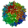
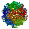
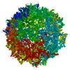
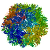
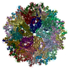
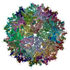
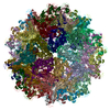
| Entry | Database: PDB / ID: 6bx0 | |||||||||||||||||||||
|---|---|---|---|---|---|---|---|---|---|---|---|---|---|---|---|---|---|---|---|---|---|---|
| Title | Atomic resolution structure of human bufavirus 2 | |||||||||||||||||||||
 Components Components | VP2 | |||||||||||||||||||||
 Keywords Keywords | VIRUS LIKE PARTICLE / parvovirus / protoparvovirus / Bufavirus | |||||||||||||||||||||
| Function / homology | Parvovirus coat protein VP2 / Parvovirus coat protein VP1/VP2 / Parvovirus coat protein VP1/VP2 / Capsid/spike protein, ssDNA virus / T=1 icosahedral viral capsid / structural molecule activity / VP2 Function and homology information Function and homology information | |||||||||||||||||||||
| Biological species |  Bufavirus-2 Bufavirus-2 | |||||||||||||||||||||
| Method | ELECTRON MICROSCOPY / single particle reconstruction / cryo EM / Resolution: 3.79 Å | |||||||||||||||||||||
 Authors Authors | Mietzsch, M. / Agbandje-McKenna, M. | |||||||||||||||||||||
 Citation Citation | Journal: Viruses / Year: 2018 Title: Atomic Resolution Structures of Human Bufaviruses Determined by Cryo-Electron Microscopy. Authors: Maria Ilyas / Mario Mietzsch / Shweta Kailasan / Elina Väisänen / Mengxiao Luo / Paul Chipman / J Kennon Smith / Justin Kurian / Duncan Sousa / Robert McKenna / Maria Söderlund-Venermo / ...Authors: Maria Ilyas / Mario Mietzsch / Shweta Kailasan / Elina Väisänen / Mengxiao Luo / Paul Chipman / J Kennon Smith / Justin Kurian / Duncan Sousa / Robert McKenna / Maria Söderlund-Venermo / Mavis Agbandje-McKenna /   Abstract: Bufavirus strain 1 (BuV1), a member of the genus of the , was first isolated from fecal samples of children with acute diarrhea in Burkina Faso. Since this initial discovery, BuVs have been isolated ...Bufavirus strain 1 (BuV1), a member of the genus of the , was first isolated from fecal samples of children with acute diarrhea in Burkina Faso. Since this initial discovery, BuVs have been isolated in several countries, including Finland, the Netherlands, and Bhutan, in pediatric patients exhibiting similar symptoms. Towards their characterization, the structures of virus-like particles of BuV1, BuV2, and BuV3, the current known genotypes, have been determined by cryo-electron microscopy and image reconstruction to 2.84, 3.79, and 3.25 Å, respectively. The BuVs, 65-73% identical in amino acid sequence, conserve the major viral protein, VP2, structure and general capsid surface features of parvoviruses. These include a core β-barrel (βB-βI), α-helix A, and large surface loops inserted between these elements in VP2. The capsid contains depressions at the icosahedral 2-fold and around the 5-fold axes, and has three separated protrusions surrounding the 3-fold axes. Structure comparison among the BuVs and to available parvovirus structures revealed capsid surface variations and capsid 3-fold protrusions that depart from the single pinwheel arrangement of the animal protoparvoviruses. These structures provide a platform to begin the molecular characterization of these potentially pathogenic viruses. | |||||||||||||||||||||
| History |
|
- Structure visualization
| Movie |
Movie viewer |
|---|---|
| Structure viewer | Molecule: MolmilJmol/JSmol |
- Downloads & links
-Download
| PDBx/mmCIF format | 6bx0.cif.gz | 5.4 MB | Display | PDBx/mmCIF format |
|---|---|---|---|---|
| PDB format | pdb6bx0.ent.gz | Display | PDB format | |
| PDBx/mmJSON format | 6bx0.json.gz | Tree view | PDBx/mmJSON format | |
| Others |  Other downloads Other downloads |
-Validation report
| Arichive directory | https://data.pdbj.org/pub/pdb/validation_reports/bx/6bx0ftp://data.pdbj.org/pub/pdb/validation_reports/bx/6bx0 | HTTPS FTP |
|---|
-Related structure data
| Related structure data |  7301MC 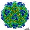 7300C  7302C  6bwxC  6bx1C M: map data used to model this data C: citing same article ( |
|---|---|
| Similar structure data |


-Links
 PDBj
PDBj


- Assembly
| Deposited unit | 
|
|---|---|
| 1 |
|
-Components
| #1: Protein | Mass: 61484.789 Da / Num. of mol.: 60 Source method: isolated from a genetically manipulated source Source: (gene. exp.) Bufavirus-2 / Production host:   Spodoptera frugiperda (fall armyworm) / References: UniProt: I6XT93 Spodoptera frugiperda (fall armyworm) / References: UniProt: I6XT93Has protein modification | Y | |
|---|
-Experimental details
-Experiment
| Experiment | Method: ELECTRON MICROSCOPY |
|---|---|
| EM experiment | Aggregation state: PARTICLE / 3D reconstruction method: single particle reconstruction |
- Sample preparation
| Component | Name: Bufavirus-2 / Type: VIRUS / Entity ID: all / Source: RECOMBINANT |
|---|---|
| Source (natural) | Organism: Bufavirus-2 |
| Source (recombinant) | Organism: Spodoptera frugiperda (fall armyworm) |
| Details of virus | Empty: YES / Enveloped: NO / Isolate: SEROTYPE / Type: VIRUS-LIKE PARTICLE |
| Natural host | Organism: Homo sapiens |
| Buffer solution | pH: 7.4 |
| Specimen | Embedding applied: NO / Shadowing applied: NO / Staining applied: NO / Vitrification applied: YES |
| Vitrification | Cryogen name: ETHANE |
- Electron microscopy imaging
| Experimental equipment |  Model: Titan Krios / Image courtesy: FEI Company |
|---|---|
| Microscopy | Model: FEI TITAN KRIOS |
| Electron gun | Electron source:  FIELD EMISSION GUN / Accelerating voltage: 300 kV / Illumination mode: FLOOD BEAM FIELD EMISSION GUN / Accelerating voltage: 300 kV / Illumination mode: FLOOD BEAM |
| Electron lens | Mode: BRIGHT FIELD / Cs: 2.7 mm |
| Image recording | Electron dose: 57 e/Å2 / Film or detector model: DIRECT ELECTRON DE-20 (5k x 3k) / Num. of real images: 429 |
| Image scans | Movie frames/image: 34 / Used frames/image: 2-20 |
- Processing
| Software | Name: PHENIX / Version: 1.10-2155_2155: / Classification: refinement | |||||||||||||||||||||||||||||||||||||||||||||
|---|---|---|---|---|---|---|---|---|---|---|---|---|---|---|---|---|---|---|---|---|---|---|---|---|---|---|---|---|---|---|---|---|---|---|---|---|---|---|---|---|---|---|---|---|---|---|
| EM software |
| |||||||||||||||||||||||||||||||||||||||||||||
| CTF correction | Type: PHASE FLIPPING AND AMPLITUDE CORRECTION | |||||||||||||||||||||||||||||||||||||||||||||
| Particle selection | Num. of particles selected: 8404 | |||||||||||||||||||||||||||||||||||||||||||||
| 3D reconstruction | Resolution: 3.79 Å / Resolution method: FSC 0.143 CUT-OFF / Num. of particles: 7564 / Symmetry type: POINT | |||||||||||||||||||||||||||||||||||||||||||||
| Atomic model building | Protocol: RIGID BODY FIT / Target criteria: Correlation coefficient | |||||||||||||||||||||||||||||||||||||||||||||
| Refine LS restraints |
|
