National Institutes of Health/National Center for Research Resources (NIH/NCRR)
R01 GM109524
United States
Citation
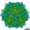
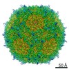
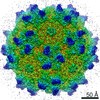
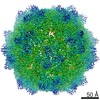
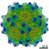
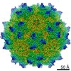
Journal: J Struct Biol / Year: 2020 Title: Structure comparison of the chimeric AAV2.7m8 vector with parental AAV2. Authors: Antonette Bennett / Annahita Keravala / Victoria Makal / Justin Kurian / Brahim Belbellaa / Rangoli Aeran / Yu-Shan Tseng / Duncan Sousa / John Spear / Mehdi Gasmi / Mavis Agbandje-McKenna / Abstract: The AAV2.7m8 vector is an engineered capsid with a 10-amino acid insertion in adeno-associated virus (AAV) surface variable region VIII (VR-VIII) resulting in the alteration of an antigenic region of ...The AAV2.7m8 vector is an engineered capsid with a 10-amino acid insertion in adeno-associated virus (AAV) surface variable region VIII (VR-VIII) resulting in the alteration of an antigenic region of AAV2 and the ability to efficiently transduce retina cells following intravitreal administration. Directed evolution and in vivo screening in the mouse retina isolated this vector. In the present study, we sought to identify the structural differences between a recombinant AAV2.7m8 (rAAV2.7m8) vector packaging a GFP genome and its parental serotype, AAV2, by cryo-electron microscopy (cryo-EM) and image reconstruction. The structures of rAAV2.7m8 and AAV2 were determined to 2.91 and 3.02 Å resolution, respectively. The rAAV2.7m8 amino acid side-chains for residues 219-745 (the last C-terminal residue) were interpretable in the density map with the exception of the 10 inserted amino acids. While observable in a low sigma threshold density, side-chains were only resolved at the base of the insertion, likely due to flexibility at the top of the loop. A comparison to parental AAV2 (ordered from residues 217-735) showed the structures to be similar, except at some side-chains that had different orientations and, in VR-VIII containing the 10 amino acid insertion. VR-VIII is part of an AAV2 antigenic epitope, and the difference is consistent with rAAV2.7m8's escape from a known AAV2 monoclonal antibody, C37-B. The observations provide valuable insight into the configuration of inserted surface peptides on the AAV capsid and structural differences to be leveraged for future AAV vector rational design, especially for retargeted tropism and antibody escape.
History
Deposition
Aug 14, 2019
-
Header (metadata) release
Sep 18, 2019
-
Map release
Aug 19, 2020
-
Update
Mar 20, 2024
-
Current status
Mar 20, 2024
Processing site: RCSB / Status: Released
-
Structure visualization
Movie
Surface view with section colored by density value
In the structure databanks used in Yorodumi, some data are registered as the other names, "COVID-19 virus" and "2019-nCoV". Here are the details of the virus and the list of structure data.
Jan 31, 2019. EMDB accession codes are about to change! (news from PDBe EMDB page)
EMDB accession codes are about to change! (news from PDBe EMDB page)
The allocation of 4 digits for EMDB accession codes will soon come to an end. Whilst these codes will remain in use, new EMDB accession codes will include an additional digit and will expand incrementally as the available range of codes is exhausted. The current 4-digit format prefixed with “EMD-” (i.e. EMD-XXXX) will advance to a 5-digit format (i.e. EMD-XXXXX), and so on. It is currently estimated that the 4-digit codes will be depleted around Spring 2019, at which point the 5-digit format will come into force.
The EM Navigator/Yorodumi systems omit the EMD- prefix.
Related info.:Q: What is EMD? / ID/Accession-code notation in Yorodumi/EM Navigator
Yorodumi is a browser for structure data from EMDB, PDB, SASBDB, etc.
This page is also the successor to EM Navigator detail page, and also detail information page/front-end page for Omokage search.
The word "yorodu" (or yorozu) is an old Japanese word meaning "ten thousand". "mi" (miru) is to see.
Related info.:EMDB / PDB / SASBDB / Comparison of 3 databanks / Yorodumi Search / Aug 31, 2016. New EM Navigator & Yorodumi / Yorodumi Papers / Jmol/JSmol / Function and homology information / Changes in new EM Navigator and Yorodumi
 Movie
Movie Controller


 Open data
 Basic information
 Map data
 Sample
 Keywords
 Function and homology information
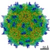
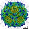
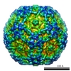
 Adeno-associated virus - 2
 Authors
 United States, 1 items
  Citation
 Structure visualization
 Downloads & links
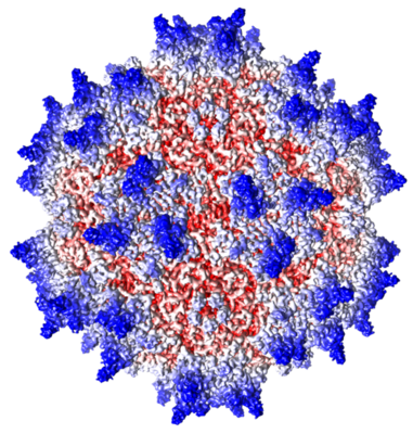 emd_20610.png
 http://ftp.pdbj.org/pub/emdb/structures/EMD-20610


 Z (Sec.)
 X (Row.)
 Y (Col.)


 Sample components
 Spodoptera frugiperda (fall armyworm)
 Processing
 Electron microscopy
 FIELD EMISSION GUN
