Mass: 18.015 Da / Num. of mol.: 971 / Source method: isolated from a natural source / Formula: H2O
-
Details
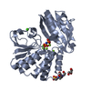
Has ligand of interest
Y
Sequence details
The cDNA sequence had a sequence coding Ser at the amino acid position of 116, which is different ...The cDNA sequence had a sequence coding Ser at the amino acid position of 116, which is different from R116 in the database entry (Uniprot L8AXY8). This conflict may be possibly derived from natural mutation or the presence of isoform.
-
Experimental details
-
Experiment
Experiment
Method: X-RAY DIFFRACTION / Number of used crystals: 1
-
Sample preparation
Crystal
Density Matthews: 2.33 Å3/Da / Density % sol: 47.21 %
Crystal grow
Temperature: 285 K / Method: vapor diffusion, sitting drop / pH: 7.4 Details: 24%(w/v) PEG3350, 10 mM ammonium phosphate dibasic and 0.2 M NaCl., 0.1 M HEPES
-
Data collection
Diffraction
Mean temperature: 100 K / Serial crystal experiment: N
Resolution: 1.3→46.568 Å / Cor.coef. Fo:Fc: 0.986 / Cor.coef. Fo:Fc free: 0.98 / SU B: 0.921 / SU ML: 0.018 / Cross valid method: THROUGHOUT / ESU R: 0.034 / ESU R Free: 0.033 Details: Hydrogens have been added in their riding positions
Rfactor
Num. reflection
% reflection
Rfree
0.1216
7540
5.054 %
Rwork
0.0965
-
-
all
0.098
-
-
obs
-
149178
98.271 %
Solvent computation
Ion probe radii: 0.8 Å / Shrinkage radii: 0.8 Å / VDW probe radii: 1.2 Å
Displacement parameters
Biso mean: 14.123 Å2
Baniso -1
Baniso -2
Baniso -3
1-
0.469 Å2
0 Å2
0.148 Å2
2-
-
-0.022 Å2
0 Å2
3-
-
-
-0.47 Å2
Refinement step
Cycle: LAST / Resolution: 1.3→46.568 Å
Protein
Nucleic acid
Ligand
Solvent
Total
Num. atoms
4680
0
85
971
5736
Refine LS restraints
Refine-ID
Type
Dev ideal
Dev ideal target
Number
X-RAY DIFFRACTION
r_bond_refined_d
0.013
0.013
5137
X-RAY DIFFRACTION
r_bond_other_d
0.001
0.017
4956
X-RAY DIFFRACTION
r_angle_refined_deg
1.796
1.648
7026
X-RAY DIFFRACTION
r_angle_other_deg
1.562
1.587
11577
X-RAY DIFFRACTION
r_dihedral_angle_1_deg
6.599
5
700
X-RAY DIFFRACTION
r_dihedral_angle_2_deg
34.083
23.644
236
X-RAY DIFFRACTION
r_dihedral_angle_3_deg
11.608
15
941
X-RAY DIFFRACTION
r_dihedral_angle_4_deg
14.566
15
24
X-RAY DIFFRACTION
r_chiral_restr
0.108
0.2
711
X-RAY DIFFRACTION
r_gen_planes_refined
0.009
0.02
5730
X-RAY DIFFRACTION
r_gen_planes_other
0.002
0.02
982
X-RAY DIFFRACTION
r_nbd_refined
0.23
0.2
1089
X-RAY DIFFRACTION
r_symmetry_nbd_other
0.179
0.2
4634
X-RAY DIFFRACTION
r_nbtor_refined
0.173
0.2
2490
X-RAY DIFFRACTION
r_symmetry_nbtor_other
0.086
0.2
2293
X-RAY DIFFRACTION
r_xyhbond_nbd_refined
0.163
0.2
675
X-RAY DIFFRACTION
r_symmetry_xyhbond_nbd_other
0.047
0.2
2
X-RAY DIFFRACTION
r_metal_ion_refined
0.015
0.2
2
X-RAY DIFFRACTION
r_symmetry_nbd_refined
0.143
0.2
10
X-RAY DIFFRACTION
r_nbd_other
0.162
0.2
50
X-RAY DIFFRACTION
r_symmetry_xyhbond_nbd_refined
0.177
0.2
46
X-RAY DIFFRACTION
r_mcbond_it
1.023
1.169
2552
X-RAY DIFFRACTION
r_mcbond_other
1.014
1.167
2551
X-RAY DIFFRACTION
r_mcangle_it
1.305
1.764
3215
X-RAY DIFFRACTION
r_mcangle_other
1.307
1.766
3216
X-RAY DIFFRACTION
r_scbond_it
1.98
1.453
2585
X-RAY DIFFRACTION
r_scbond_other
1.98
1.454
2586
X-RAY DIFFRACTION
r_scangle_it
2.37
2.075
3768
X-RAY DIFFRACTION
r_scangle_other
2.37
2.076
3769
X-RAY DIFFRACTION
r_lrange_it
2.925
16.755
6207
X-RAY DIFFRACTION
r_lrange_other
2.691
16.083
6057
X-RAY DIFFRACTION
r_rigid_bond_restr
2.249
3
10093
LS refinement shell
Resolution (Å)
Rfactor Rfree
Num. reflection Rfree
Rfactor Rwork
Num. reflection Rwork
Refine-ID
% reflection obs (%)
1.3-1.334
0.209
430
0.183
8611
X-RAY DIFFRACTION
81.1871
1.334-1.37
0.163
525
0.134
9988
X-RAY DIFFRACTION
95.8865
1.37-1.41
0.134
548
0.099
10020
X-RAY DIFFRACTION
99.83
1.41-1.453
0.114
498
0.077
9800
X-RAY DIFFRACTION
99.9903
1.453-1.501
0.104
519
0.068
9455
X-RAY DIFFRACTION
100
1.501-1.554
0.092
515
0.062
9154
X-RAY DIFFRACTION
100
1.554-1.612
0.105
469
0.065
8858
X-RAY DIFFRACTION
100
1.612-1.678
0.102
460
0.065
8558
X-RAY DIFFRACTION
100
1.678-1.753
0.104
400
0.069
8172
X-RAY DIFFRACTION
99.9883
1.753-1.838
0.108
426
0.07
7852
X-RAY DIFFRACTION
100
1.838-1.938
0.108
415
0.078
7384
X-RAY DIFFRACTION
100
1.938-2.055
0.102
383
0.083
7086
X-RAY DIFFRACTION
99.9866
2.055-2.197
0.113
340
0.084
6604
X-RAY DIFFRACTION
100
2.197-2.373
0.107
346
0.086
6146
X-RAY DIFFRACTION
99.9846
2.373-2.599
0.113
297
0.092
5706
X-RAY DIFFRACTION
100
2.599-2.905
0.131
273
0.105
5138
X-RAY DIFFRACTION
99.9815
2.905-3.354
0.139
242
0.113
4549
X-RAY DIFFRACTION
100
3.354-4.105
0.132
192
0.11
3877
X-RAY DIFFRACTION
99.9754
4.105-5.797
0.142
168
0.127
2998
X-RAY DIFFRACTION
99.9369
5.797-46.568
0.163
94
0.184
1682
X-RAY DIFFRACTION
99.8314
+
About Yorodumi
-
News
-
Feb 9, 2022. New format data for meta-information of EMDB entries
New format data for meta-information of EMDB entries
Version 3 of the EMDB header file is now the official format.
The previous official version 1.9 will be removed from the archive.
In the structure databanks used in Yorodumi, some data are registered as the other names, "COVID-19 virus" and "2019-nCoV". Here are the details of the virus and the list of structure data.
Jan 31, 2019. EMDB accession codes are about to change! (news from PDBe EMDB page)
EMDB accession codes are about to change! (news from PDBe EMDB page)
The allocation of 4 digits for EMDB accession codes will soon come to an end. Whilst these codes will remain in use, new EMDB accession codes will include an additional digit and will expand incrementally as the available range of codes is exhausted. The current 4-digit format prefixed with “EMD-” (i.e. EMD-XXXX) will advance to a 5-digit format (i.e. EMD-XXXXX), and so on. It is currently estimated that the 4-digit codes will be depleted around Spring 2019, at which point the 5-digit format will come into force.
The EM Navigator/Yorodumi systems omit the EMD- prefix.
Related info.:Q: What is EMD? / ID/Accession-code notation in Yorodumi/EM Navigator
Yorodumi is a browser for structure data from EMDB, PDB, SASBDB, etc.
This page is also the successor to EM Navigator detail page, and also detail information page/front-end page for Omokage search.
The word "yorodu" (or yorozu) is an old Japanese word meaning "ten thousand". "mi" (miru) is to see.
Related info.:EMDB / PDB / SASBDB / Comparison of 3 databanks / Yorodumi Search / Aug 31, 2016. New EM Navigator & Yorodumi / Yorodumi Papers / Jmol/JSmol / Function and homology information / Changes in new EM Navigator and Yorodumi
 Movie
Movie Controller

 Yorodumi
 Open data
 Basic information
 Components
 Keywords
 Function and homology information
 Shewanella sp. AS-11 (bacteria)
 X-RAY DIFFRACTION /
  Authors
 Japan, 2items
  Citation
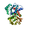
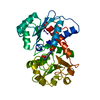
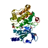
 Structure visualization
 Downloads & links
 Other downloads
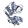


 PDBj
PDBj Assembly
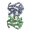


 Mass: 24.305 Da / Num. of mol.: 10 / Source method: obtained synthetically / Formula: Mg
 Mass: 40.078 Da / Num. of mol.: 1 / Source method: obtained synthetically / Formula: Ca
 Mass: 176.990 Da / Num. of mol.: 2 / Source method: obtained synthetically / Formula: H5NO6P2 / Feature type: SUBJECT OF INVESTIGATION
 Mass: 238.305 Da / Num. of mol.: 2 / Source method: obtained synthetically / Formula: C8H18N2O4S / Comment: pH buffer*YM
 Mass: 92.094 Da / Num. of mol.: 4 / Source method: obtained synthetically / Formula: C3H8O3
 Mass: 18.998 Da / Num. of mol.: 2 / Source method: obtained synthetically / Formula: F
 Sample preparation
 Processing