 Movie
Movie Controller

[English] 日本語
 Yorodumi
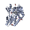
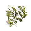
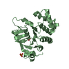
- PDB-7c7m: The structure of SAM-bound CntL, an aminobutyrate transferase in ... -
+ Open data

- Basic information
| Entry | Database: PDB / ID: 7c7m | ||||||
|---|---|---|---|---|---|---|---|
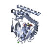
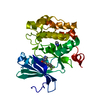
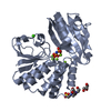
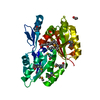
| Title | The structure of SAM-bound CntL, an aminobutyrate transferase in staphylopine biosysnthesis | ||||||
 Components Components | Staphylopine biosynthesis enzyme CntL | ||||||
 Keywords Keywords | TRANSFERASE / Metallophore / Biosynthesis / Substrate / Aminobutyrate | ||||||
| Function / homology | nicotianamine synthase activity / nicotianamine biosynthetic process / Nicotianamine synthase / methyltransferase activity / methylation / S-adenosyl-L-methionine-dependent methyltransferase superfamily / S-ADENOSYLMETHIONINE / D-histidine (S)-2-aminobutanoyltransferase CntL Function and homology information Function and homology information | ||||||
| Biological species |   Staphylococcus aureus (bacteria) Staphylococcus aureus (bacteria) | ||||||
| Method |  X-RAY DIFFRACTION / SYNCHROTRON / SAD / Resolution: 1.81 Å X-RAY DIFFRACTION / SYNCHROTRON / SAD / Resolution: 1.81 Å | ||||||
 Authors Authors | Luo, Z. / Luo, S. / Zhou, H. | ||||||
| Funding support |  China, 1items China, 1items
| ||||||
 Citation Citation | Journal: Faseb J. / Year: 2021 Title: Structural insights into the ligand recognition and catalysis of the key aminobutanoyltransferase CntL in staphylopine biosynthesis. Authors: Luo, Z. / Luo, S. / Ju, Y. / Ding, P. / Xu, J. / Gu, Q. / Zhou, H. | ||||||
| History |
|
- Structure visualization
| Structure viewer | Molecule: MolmilJmol/JSmol |
|---|
- Downloads & links
-Download
| PDBx/mmCIF format | 7c7m.cif.gz | 72.4 KB | Display | PDBx/mmCIF format |
|---|---|---|---|---|
| PDB format | pdb7c7m.ent.gz | 50.6 KB | Display | PDB format |
| PDBx/mmJSON format | 7c7m.json.gz | Tree view | PDBx/mmJSON format | |
| Others |  Other downloads Other downloads |
-Validation report
| Arichive directory | https://data.pdbj.org/pub/pdb/validation_reports/c7/7c7mftp://data.pdbj.org/pub/pdb/validation_reports/c7/7c7m | HTTPS FTP |
|---|
-Related structure data


-Links
 PDBj
PDBj
- Assembly
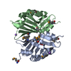
| Deposited unit | 
| ||||||||||||
|---|---|---|---|---|---|---|---|---|---|---|---|---|---|
| 1 |
| ||||||||||||
| Unit cell |
| ||||||||||||
| Components on special symmetry positions |
|
-Components
| #1: Protein | Mass: 32582.947 Da / Num. of mol.: 1 / Mutation: A119T Source method: isolated from a genetically manipulated source Details: M(INITIATING METHIONINE)GSSHHHHHHSS G(EXPRESSION TAG) Source: (gene. exp.) Staphylococcus aureus (bacteria) / Gene: fmrO, cntL, DDL17_02690, M1K003_1012Production host: References: UniProt: A0A391FC11 | ||||||||
|---|---|---|---|---|---|---|---|---|---|
| #2: Chemical |   Mass: 62.068 Da / Num. of mol.: 2 / Source method: obtained synthetically / Formula: C2H6O2 / Feature type: SUBJECT OF INVESTIGATION Mass: 62.068 Da / Num. of mol.: 2 / Source method: obtained synthetically / Formula: C2H6O2 / Feature type: SUBJECT OF INVESTIGATION#3: Chemical | ChemComp-SAM / |   Mass: 398.437 Da / Num. of mol.: 1 / Source method: isolated from a natural source / Formula: C15H22N6O5S / Feature type: SUBJECT OF INVESTIGATION Mass: 398.437 Da / Num. of mol.: 1 / Source method: isolated from a natural source / Formula: C15H22N6O5S / Feature type: SUBJECT OF INVESTIGATION#4: Water | ChemComp-HOH / |  Mass: 18.015 Da / Num. of mol.: 153 / Source method: isolated from a natural source / Formula: H2O Mass: 18.015 Da / Num. of mol.: 153 / Source method: isolated from a natural source / Formula: H2OHas ligand of interest | Y | Has protein modification | Y | |
-Experimental details
-Experiment
| Experiment | Method: X-RAY DIFFRACTION / Number of used crystals: 1 |
|---|
- Sample preparation
| Crystal | Density Matthews: 1.82 Å3/Da / Density % sol: 32.45 % |
|---|---|
| Crystal grow | Temperature: 291 K / Method: vapor diffusion, sitting drop / Details: 0.1 M Bis-Tris pH 5.5, 2.0 M Ammonium sulfate |
-Data collection
| Diffraction | Mean temperature: 100 K / Serial crystal experiment: N |
|---|---|
| Diffraction source | Source: SYNCHROTRON / Site: SSRF / Beamline: BL19U1 / Wavelength: 1 Å |
| Detector | Type: DECTRIS PILATUS3 6M / Detector: PIXEL / Date: Dec 10, 2018 |
| Radiation | Protocol: SINGLE WAVELENGTH / Monochromatic (M) / Laue (L): M / Scattering type: x-ray |
| Radiation wavelength | Wavelength: 1 Å / Relative weight: 1 |
| Reflection | Resolution: 1.81→48.27 Å / Num. obs: 22386 / % possible obs: 99.8 % / Redundancy: 12.8 % / CC1/2: 0.999 / Rmerge(I) obs: 0.088 / Net I/σ(I): 21.4 |
| Reflection shell | Resolution: 1.81→1.85 Å / Rmerge(I) obs: 0.56 / Num. unique obs: 1292 / CC1/2: 0.958 |
- Processing
| Software |
| ||||||||||||||||||||||||||||||||||||||||||||||||||||||||||||||||||||||||||||||||||||||||||||||||||||||||||||||||||||||||||||||||||||||||||||||||||||||||||||||||||||||||||||||||||||||
|---|---|---|---|---|---|---|---|---|---|---|---|---|---|---|---|---|---|---|---|---|---|---|---|---|---|---|---|---|---|---|---|---|---|---|---|---|---|---|---|---|---|---|---|---|---|---|---|---|---|---|---|---|---|---|---|---|---|---|---|---|---|---|---|---|---|---|---|---|---|---|---|---|---|---|---|---|---|---|---|---|---|---|---|---|---|---|---|---|---|---|---|---|---|---|---|---|---|---|---|---|---|---|---|---|---|---|---|---|---|---|---|---|---|---|---|---|---|---|---|---|---|---|---|---|---|---|---|---|---|---|---|---|---|---|---|---|---|---|---|---|---|---|---|---|---|---|---|---|---|---|---|---|---|---|---|---|---|---|---|---|---|---|---|---|---|---|---|---|---|---|---|---|---|---|---|---|---|---|---|---|---|---|---|
| Refinement | Method to determine structure: SAD / Resolution: 1.81→48.27 Å / Cor.coef. Fo:Fc: 0.953 / Cor.coef. Fo:Fc free: 0.949 / SU B: 2.726 / SU ML: 0.083 / Cross valid method: THROUGHOUT / ESU R: 0.145 / ESU R Free: 0.123 / Stereochemistry target values: MAXIMUM LIKELIHOOD / Details: HYDROGENS HAVE BEEN ADDED IN THE RIDING POSITIONS
| ||||||||||||||||||||||||||||||||||||||||||||||||||||||||||||||||||||||||||||||||||||||||||||||||||||||||||||||||||||||||||||||||||||||||||||||||||||||||||||||||||||||||||||||||||||||
| Solvent computation | Ion probe radii: 0.8 Å / Shrinkage radii: 0.8 Å / VDW probe radii: 1.2 Å / Solvent model: MASK | ||||||||||||||||||||||||||||||||||||||||||||||||||||||||||||||||||||||||||||||||||||||||||||||||||||||||||||||||||||||||||||||||||||||||||||||||||||||||||||||||||||||||||||||||||||||
| Displacement parameters | Biso mean: 20.593 Å2
| ||||||||||||||||||||||||||||||||||||||||||||||||||||||||||||||||||||||||||||||||||||||||||||||||||||||||||||||||||||||||||||||||||||||||||||||||||||||||||||||||||||||||||||||||||||||
| Refinement step | Cycle: 1 / Resolution: 1.81→48.27 Å
| ||||||||||||||||||||||||||||||||||||||||||||||||||||||||||||||||||||||||||||||||||||||||||||||||||||||||||||||||||||||||||||||||||||||||||||||||||||||||||||||||||||||||||||||||||||||
| Refine LS restraints |
|