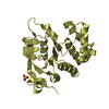
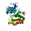
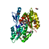
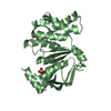
- PDB-3grd: Crystal structure of NTF2-superfamily protein with unknown functi... -
+
Open data
ID or keywords:
Loading...
-
Basic information
Entry
Database: PDB / ID: 3grd
Title
Crystal structure of NTF2-superfamily protein with unknown function (NP_977240.1) from BACILLUS CEREUS ATCC 10987 at 1.25 A resolution
Components
uncharacterized NTF2-superfamily protein
Keywords
structural genomics / unknown function / NP_977240.1 / NTF2-superfamily protein with unknown function / Joint Center for Structural Genomics / JCSG / Protein Structure Initiative / PSI-2
Function / homology
SnoaL-like domain / SnoaL-like domain / Nuclear Transport Factor 2; Chain: A, - #50 / NTF2-like domain superfamily / Nuclear Transport Factor 2; Chain: A, / Roll / Alpha Beta / ACETATE ION / SnoaL-like domain-containing protein
Function and homology information
Biological species
Bacillus cereus ATCC 10987 (bacteria)
Method
X-RAY DIFFRACTION / SYNCHROTRON / MAD / Resolution: 1.25 Å
Mass: 18.015 Da / Num. of mol.: 521 / Source method: isolated from a natural source / Formula: H2O
Has protein modification
Y
Sequence details
THE CONSTRUCT WAS EXPRESSED WITH A PURIFICATION TAG MGSDKIHHHHHHENLYFQG. THE TAG WAS REMOVED WITH ...THE CONSTRUCT WAS EXPRESSED WITH A PURIFICATION TAG MGSDKIHHHHHHENLYFQG. THE TAG WAS REMOVED WITH TEV PROTEASE LEAVING ONLY A GLYCINE (0) FOLLOWED BY THE TARGET SEQUENCE.
-
Experimental details
-
Experiment
Experiment
Method: X-RAY DIFFRACTION / Number of used crystals: 1
-
Sample preparation
Crystal
Density Matthews: 2.41 Å3/Da / Density % sol: 48.87 %
Crystal grow
Temperature: 277 K / Method: vapor diffusion, sitting drop / pH: 8.29 Details: 33.8000% polyethylene glycol 4000, 0.2000M sodium acetate, 0.1M TRIS pH 8.29, NANODROP, VAPOR DIFFUSION, SITTING DROP, temperature 277K
Type: MARMOSAIC 325 mm CCD / Detector: CCD / Date: Nov 15, 2008 / Details: Flat mirror (vertical focusing)
Radiation
Monochromator: Single crystal Si(111) bent monochromator (horizontal focusing) Protocol: MAD / Monochromatic (M) / Laue (L): M / Scattering type: x-ray
Radiation wavelength
ID
Wavelength (Å)
Relative weight
1
0.91162
1
2
0.97862
1
Reflection
Resolution: 1.25→29.566 Å / Num. obs: 81747 / % possible obs: 96.1 % / Observed criterion σ(I): -3 / Biso Wilson estimate: 10.01 Å2 / Rmerge(I) obs: 0.041 / Net I/σ(I): 11.63
Reflection shell
Resolution (Å)
Rmerge(I) obs
Mean I/σ(I) obs
Num. measured obs
Num. unique obs
% possible all
1.25-1.29
0.469
1.6
20216
11671
82.5
1.29-1.35
0.387
2
30846
17428
95.2
1.35-1.41
0.301
2.6
26447
14719
96
1.41-1.48
0.229
3.4
26004
14313
96.4
1.48-1.57
0.151
5.1
27599
15005
97.1
1.57-1.7
0.11
7.1
30794
16539
98.1
1.7-1.87
0.068
10.8
29117
15423
98.7
1.87-2.14
0.037
18.2
29546
15469
98.8
2.14-2.69
0.026
25.3
29715
15426
98.8
2.69-29.566
0.017
37.8
30284
15579
98.2
-
Phasing
Phasing
Method: MAD
-
Processing
Software
Name
Version
Classification
NB
REFMAC
5.2.0019
refinement
PHENIX
refinement
SHELX
phasing
MolProbity
3beta29
modelbuilding
XSCALE
datascaling
PDB_EXTRACT
3.006
dataextraction
XDS
datareduction
SHELXD
phasing
autoSHARP
phasing
Refinement
Method to determine structure: MAD / Resolution: 1.25→29.566 Å / Cor.coef. Fo:Fc: 0.977 / Cor.coef. Fo:Fc free: 0.968 / Occupancy max: 1 / Occupancy min: 0.1 / SU B: 1.289 / SU ML: 0.025 / Cross valid method: THROUGHOUT / σ(F): 0 / ESU R: 0.04 / ESU R Free: 0.041 Stereochemistry target values: MAXIMUM LIKELIHOOD WITH PHASES Details: 1. HYDROGENS HAVE BEEN ADDED IN THE RIDING POSITIONS. 2. A MET-INHIBITION PROTOCOL WAS USED FOR SELENOMETHIONINE INCORPORATION DURING PROTEIN EXPRESSION. THE OCCUPANCY OF THE SE ATOMS IN THE ...Details: 1. HYDROGENS HAVE BEEN ADDED IN THE RIDING POSITIONS. 2. A MET-INHIBITION PROTOCOL WAS USED FOR SELENOMETHIONINE INCORPORATION DURING PROTEIN EXPRESSION. THE OCCUPANCY OF THE SE ATOMS IN THE MSE RESIDUES WAS REDUCED TO 0.75 FOR THE REDUCED SCATTERING POWER DUE TO PARTIAL S-MET INCORPORATION. 3. ACETATE (ACT) AND SODIUM (NA) IONS FROM CRYSTALLIZATION CONDITION ARE MODELED.
Rfactor
Num. reflection
% reflection
Selection details
Rfree
0.167
4091
5 %
RANDOM
Rwork
0.133
-
-
-
obs
0.135
81659
98.96 %
-
Solvent computation
Ion probe radii: 0.8 Å / Shrinkage radii: 0.8 Å / VDW probe radii: 1.2 Å / Solvent model: MASK
In the structure databanks used in Yorodumi, some data are registered as the other names, "COVID-19 virus" and "2019-nCoV". Here are the details of the virus and the list of structure data.
Jan 31, 2019. EMDB accession codes are about to change! (news from PDBe EMDB page)
EMDB accession codes are about to change! (news from PDBe EMDB page)
The allocation of 4 digits for EMDB accession codes will soon come to an end. Whilst these codes will remain in use, new EMDB accession codes will include an additional digit and will expand incrementally as the available range of codes is exhausted. The current 4-digit format prefixed with “EMD-” (i.e. EMD-XXXX) will advance to a 5-digit format (i.e. EMD-XXXXX), and so on. It is currently estimated that the 4-digit codes will be depleted around Spring 2019, at which point the 5-digit format will come into force.
The EM Navigator/Yorodumi systems omit the EMD- prefix.
Related info.:Q: What is EMD? / ID/Accession-code notation in Yorodumi/EM Navigator
Yorodumi is a browser for structure data from EMDB, PDB, SASBDB, etc.
This page is also the successor to EM Navigator detail page, and also detail information page/front-end page for Omokage search.
The word "yorodu" (or yorozu) is an old Japanese word meaning "ten thousand". "mi" (miru) is to see.
Related info.:EMDB / PDB / SASBDB / Comparison of 3 databanks / Yorodumi Search / Aug 31, 2016. New EM Navigator & Yorodumi / Yorodumi Papers / Jmol/JSmol / Function and homology information / Changes in new EM Navigator and Yorodumi
 Movie
Movie Controller

 Yorodumi
 Open data
 Basic information
 Components
 Keywords
 Function and homology information
 X-RAY DIFFRACTION /
  Authors
 Citation
 Structure visualization
 Downloads & links
 Other downloads
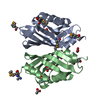


 PDBj
PDBj
 Assembly
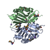

 Mass: 59.044 Da / Num. of mol.: 2 / Source method: obtained synthetically / Formula: C2H3O2
 Mass: 22.990 Da / Num. of mol.: 2 / Source method: obtained synthetically / Formula: Na
 Mass: 18.015 Da / Num. of mol.: 521 / Source method: isolated from a natural source / Formula: H2O
 Sample preparation
 / Beamline: BL11-1 / Wavelength: 0.91162,0.97862
 Processing