 Movie
Movie Controller

[English] 日本語
 Yorodumi
- PDB-1oth: CRYSTAL STRUCTURE OF HUMAN ORNITHINE TRANSCARBAMOYLASE COMPLEXED ... -
+ Open data

- Basic information
| Entry | Database: PDB / ID: 1oth | |||||||||
|---|---|---|---|---|---|---|---|---|---|---|
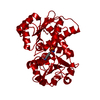
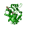
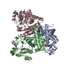
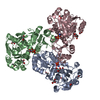
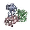
| Title | CRYSTAL STRUCTURE OF HUMAN ORNITHINE TRANSCARBAMOYLASE COMPLEXED WITH N-PHOSPHONACETYL-L-ORNITHINE | |||||||||
 Components Components | PROTEIN (ORNITHINE TRANSCARBAMOYLASE) | |||||||||
 Keywords Keywords | TRANSFERASE / TRANSCARBAMOYLASE | |||||||||
| Function / homology |  Function and homology information Function and homology informationresponse to biotin / L-ornithine catabolic process / monoatomic anion homeostasis / ammonium homeostasis / ornithine carbamoyltransferase / ornithine carbamoyltransferase activity / midgut development / Urea cycle / L-citrulline biosynthetic process / : ...response to biotin / L-ornithine catabolic process / monoatomic anion homeostasis / ammonium homeostasis / ornithine carbamoyltransferase / ornithine carbamoyltransferase activity / midgut development / Urea cycle / L-citrulline biosynthetic process / : / urea cycle / Mitochondrial protein import / response to zinc ion / amino acid binding / phosphate ion binding / liver development / phospholipid binding / response to insulin / mitochondrial inner membrane / mitochondrial matrix / response to xenobiotic stimulus / mitochondrion / identical protein binding Similarity search - Function | |||||||||
| Biological species |  Homo sapiens (human) Homo sapiens (human) | |||||||||
| Method |  X-RAY DIFFRACTION / SYNCHROTRON / MOLECULAR REPLACEMENT / Resolution: 1.85 Å X-RAY DIFFRACTION / SYNCHROTRON / MOLECULAR REPLACEMENT / Resolution: 1.85 Å | |||||||||
 Authors Authors | Shi, D. / Morizono, H. / Ha, Y. / Aoyagi, M. / Tuchman, N. / Allewell, N.M. | |||||||||
 Citation Citation | Journal: J.Biol.Chem. / Year: 1998 Title: 1.85-A resolution crystal structure of human ornithine transcarbamoylase complexed with N-phosphonacetyl-L-ornithine. Catalytic mechanism and correlation with inherited deficiency. Authors: Shi, D. / Morizono, H. / Ha, Y. / Aoyagi, M. / Tuchman, M. / Allewell, N.M. | |||||||||
| History |
|
- Structure visualization
| Structure viewer | Molecule: MolmilJmol/JSmol |
|---|
- Downloads & links
-Download
| PDBx/mmCIF format | 1oth.cif.gz | 82.4 KB | Display | PDBx/mmCIF format |
|---|---|---|---|---|
| PDB format | pdb1oth.ent.gz | 59.6 KB | Display | PDB format |
| PDBx/mmJSON format | 1oth.json.gz | Tree view | PDBx/mmJSON format | |
| Others |  Other downloads Other downloads |
-Validation report
| Arichive directory | https://data.pdbj.org/pub/pdb/validation_reports/ot/1othftp://data.pdbj.org/pub/pdb/validation_reports/ot/1oth | HTTPS FTP |
|---|
-Related structure data
| Related structure data | 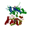 2ortS S: Starting model for refinement |
|---|---|
| Similar structure data |

-Links
 PDBj
PDBj
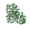
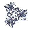
- Assembly
| Deposited unit | 
| ||||||||
|---|---|---|---|---|---|---|---|---|---|
| 1 | 
| ||||||||
| Unit cell |
| ||||||||
| Components on special symmetry positions |
|
-Components
| #1: Protein | Mass: 36106.551 Da / Num. of mol.: 1 Source method: isolated from a genetically manipulated source Details: N-PHOSPHONACETYL-L-ORNITHINE BIND ACTIVE SITE / Source: (gene. exp.) Homo sapiens (human) / Plasmid: PETOTCII / Production host:  |
|---|---|
| #2: Chemical | ChemComp-PAO /   Mass: 254.178 Da / Num. of mol.: 1 / Source method: obtained synthetically / Formula: C7H15N2O6P Mass: 254.178 Da / Num. of mol.: 1 / Source method: obtained synthetically / Formula: C7H15N2O6P |
| #3: Water | ChemComp-HOH /  Mass: 18.015 Da / Num. of mol.: 215 / Source method: isolated from a natural source / Formula: H2O Mass: 18.015 Da / Num. of mol.: 215 / Source method: isolated from a natural source / Formula: H2O |
-Experimental details
-Experiment
| Experiment | Method: X-RAY DIFFRACTION / Number of used crystals: 1 |
|---|
- Sample preparation
| Crystal | Density Matthews: 4.8 Å3/Da / Density % sol: 73 % | ||||||||||||||||||||||||||||||||||||||||||||||||||||||
|---|---|---|---|---|---|---|---|---|---|---|---|---|---|---|---|---|---|---|---|---|---|---|---|---|---|---|---|---|---|---|---|---|---|---|---|---|---|---|---|---|---|---|---|---|---|---|---|---|---|---|---|---|---|---|---|
| Crystal grow | pH: 7.5 Details: 20 MM TRISAC, 2MM EDTA, 20MM KCL, 4MM PALO PH=7.4, pH 7.5 | ||||||||||||||||||||||||||||||||||||||||||||||||||||||
| Crystal grow | *PLUS Temperature: 18 ℃ / pH: 7.4 / Method: vapor diffusion, hanging drop | ||||||||||||||||||||||||||||||||||||||||||||||||||||||
| Components of the solutions | *PLUS
|
-Data collection
| Diffraction | Mean temperature: 120 K |
|---|---|
| Diffraction source | Source: SYNCHROTRON / Site: NSLS  / Beamline: X12C / Wavelength: 1.1 / Beamline: X12C / Wavelength: 1.1 |
| Detector | Date: Mar 15, 1998 |
| Radiation | Protocol: SINGLE WAVELENGTH / Monochromatic (M) / Laue (L): M / Scattering type: x-ray |
| Radiation wavelength | Wavelength: 1.1 Å / Relative weight: 1 |
| Reflection | Resolution: 1.85→40 Å / Num. obs: 51424 / % possible obs: 86.4 % / Observed criterion σ(I): 1 / Redundancy: 28 % / Biso Wilson estimate: 15.7 Å2 / Rmerge(I) obs: 0.093 / Net I/σ(I): 12.2 |
| Reflection shell | Resolution: 1.85→1.93 Å / Redundancy: 25 % / Rmerge(I) obs: 0.27 / Mean I/σ(I) obs: 3.4 / % possible all: 63.3 |
| Reflection | *PLUS Num. measured all: 1393080 |
| Reflection shell | *PLUS % possible obs: 63.3 % / Num. unique obs: 51424 / Rmerge(I) obs: 0.27 |
- Processing
| Software |
| ||||||||||||||||||||||||||||||||||||||||||||||||||||||||||||
|---|---|---|---|---|---|---|---|---|---|---|---|---|---|---|---|---|---|---|---|---|---|---|---|---|---|---|---|---|---|---|---|---|---|---|---|---|---|---|---|---|---|---|---|---|---|---|---|---|---|---|---|---|---|---|---|---|---|---|---|---|---|
| Refinement | Method to determine structure: MOLECULAR REPLACEMENT Starting model: PDB ENTRY 2ORT Resolution: 1.85→40 Å / Data cutoff high absF: 10000000 / Data cutoff low absF: 0.001 / Cross valid method: THROUGHOUT / σ(F): 2
| ||||||||||||||||||||||||||||||||||||||||||||||||||||||||||||
| Displacement parameters | Biso mean: 17.8 Å2 | ||||||||||||||||||||||||||||||||||||||||||||||||||||||||||||
| Refinement step | Cycle: LAST / Resolution: 1.85→40 Å
| ||||||||||||||||||||||||||||||||||||||||||||||||||||||||||||
| Refine LS restraints |
| ||||||||||||||||||||||||||||||||||||||||||||||||||||||||||||
| LS refinement shell | Resolution: 1.85→1.93 Å / Total num. of bins used: 8
| ||||||||||||||||||||||||||||||||||||||||||||||||||||||||||||
| Xplor file | Serial no: 1 / Param file: PARAM19X.PRO / Topol file: TOPH19X.PRO | ||||||||||||||||||||||||||||||||||||||||||||||||||||||||||||
| Software | *PLUS Name: X-PLOR / Version: 3.851 / Classification: refinement | ||||||||||||||||||||||||||||||||||||||||||||||||||||||||||||
| Refinement | *PLUS Lowest resolution: 40 Å / σ(F): 2 / % reflection Rfree: 10 % | ||||||||||||||||||||||||||||||||||||||||||||||||||||||||||||
| Solvent computation | *PLUS | ||||||||||||||||||||||||||||||||||||||||||||||||||||||||||||
| Displacement parameters | *PLUS Biso mean: 17.8 Å2 | ||||||||||||||||||||||||||||||||||||||||||||||||||||||||||||
| Refine LS restraints | *PLUS
| ||||||||||||||||||||||||||||||||||||||||||||||||||||||||||||
| LS refinement shell | *PLUS Rfactor Rfree: 0.251 / % reflection Rfree: 8.6 % / Rfactor Rwork: 0.243 / Rfactor obs: 0.243 |
