 Movie
Movie Controller

[English] 日本語
 Yorodumi
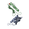
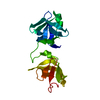
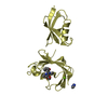
- PDB-5xby: Crystal structure of the PKA-Protein A fusion protein (end-to-end... -
+ Open data

- Basic information
| Entry | Database: PDB / ID: 5xby | ||||||
|---|---|---|---|---|---|---|---|
| Title | Crystal structure of the PKA-Protein A fusion protein (end-to-end fusion) | ||||||
 Components Components | cAMP-dependent protein kinase type II-alpha regulatory subunit,Immunoglobulin G-binding protein A | ||||||
 Keywords Keywords | SIGNALING PROTEIN / PROTEIN BINDING / synthetic protein / kinase / LYASE | ||||||
| Function / homology |  Function and homology information Function and homology informationROBO receptors bind AKAP5 / cAMP-dependent protein kinase regulator activity / nucleotide-activated protein kinase complex / cAMP-dependent protein kinase inhibitor activity / IgG binding / cAMP-dependent protein kinase complex / CREB1 phosphorylation through the activation of Adenylate Cyclase / PKA activation / ciliary base / protein kinase A catalytic subunit binding ...ROBO receptors bind AKAP5 / cAMP-dependent protein kinase regulator activity / nucleotide-activated protein kinase complex / cAMP-dependent protein kinase inhibitor activity / IgG binding / cAMP-dependent protein kinase complex / CREB1 phosphorylation through the activation of Adenylate Cyclase / PKA activation / ciliary base / protein kinase A catalytic subunit binding / PKA activation in glucagon signalling / plasma membrane raft / DARPP-32 events / cAMP binding / vascular endothelial cell response to laminar fluid shear stress / renal water homeostasis / negative regulation of cAMP/PKA signal transduction / Hedgehog 'off' state / cellular response to glucagon stimulus / FCGR3A-mediated IL10 synthesis / negative regulation of inflammatory response to antigenic stimulus / Vasopressin regulates renal water homeostasis via Aquaporins / Glucagon-like Peptide-1 (GLP1) regulates insulin secretion / ADORA2B mediated anti-inflammatory cytokines production / GPER1 signaling / Factors involved in megakaryocyte development and platelet production / adenylate cyclase-activating G protein-coupled receptor signaling pathway / High laminar flow shear stress activates signaling by PIEZO1 and PECAM1:CDH5:KDR in endothelial cells / chemical synaptic transmission / intracellular signal transduction / protein domain specific binding / focal adhesion / ubiquitin protein ligase binding / centrosome / synapse / protein-containing complex / extracellular exosome / membrane / plasma membrane / cytoplasm / cytosol Similarity search - Function | ||||||
| Biological species |  Homo sapiens (human) Homo sapiens (human)  Staphylococcus aureus (bacteria) Staphylococcus aureus (bacteria) | ||||||
| Method |  X-RAY DIFFRACTION / SYNCHROTRON / MOLECULAR REPLACEMENT / Resolution: 3.25 Å X-RAY DIFFRACTION / SYNCHROTRON / MOLECULAR REPLACEMENT / Resolution: 3.25 Å | ||||||
 Authors Authors | Youn, S.J. / Kwon, N.Y. / Lee, J.H. / Kim, J.H. / Lee, H. / Lee, J.O. | ||||||
 Citation Citation | Journal: Sci Rep / Year: 2017 Title: Construction of novel repeat proteins with rigid and predictable structures using a shared helix method. Authors: Youn, S.J. / Kwon, N.Y. / Lee, J.H. / Kim, J.H. / Choi, J. / Lee, H. / Lee, J.O. | ||||||
| History |
|
- Structure visualization
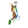
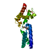
| Structure viewer | Molecule: MolmilJmol/JSmol |
|---|
- Downloads & links
-Download
| PDBx/mmCIF format | 5xby.cif.gz | 143.9 KB | Display | PDBx/mmCIF format |
|---|---|---|---|---|
| PDB format | pdb5xby.ent.gz | 116.9 KB | Display | PDB format |
| PDBx/mmJSON format | 5xby.json.gz | Tree view | PDBx/mmJSON format | |
| Others |  Other downloads Other downloads |
-Validation report
| Arichive directory | https://data.pdbj.org/pub/pdb/validation_reports/xb/5xbyftp://data.pdbj.org/pub/pdb/validation_reports/xb/5xby | HTTPS FTP |
|---|
-Related structure data
| Related structure data |  5h75C  5h76C 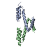 5h77C 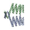 5h78C  5h79C  5h7aC  5h7bC  5h7cC  5h7dC  5x3fC  2izxS  4zncS S: Starting model for refinement C: citing same article ( |
|---|---|
| Similar structure data |


-Links
 PDBj
PDBj


- Assembly
| Deposited unit | 
| ||||||||||||||||||||||||||||||||||||||||||||||||||||||||||||||||||||||||||||||||||||||||||||||||||||||||||||||||||||||||||||||||||||||||||||||||||||||||||||||||||||||||||||||||
|---|---|---|---|---|---|---|---|---|---|---|---|---|---|---|---|---|---|---|---|---|---|---|---|---|---|---|---|---|---|---|---|---|---|---|---|---|---|---|---|---|---|---|---|---|---|---|---|---|---|---|---|---|---|---|---|---|---|---|---|---|---|---|---|---|---|---|---|---|---|---|---|---|---|---|---|---|---|---|---|---|---|---|---|---|---|---|---|---|---|---|---|---|---|---|---|---|---|---|---|---|---|---|---|---|---|---|---|---|---|---|---|---|---|---|---|---|---|---|---|---|---|---|---|---|---|---|---|---|---|---|---|---|---|---|---|---|---|---|---|---|---|---|---|---|---|---|---|---|---|---|---|---|---|---|---|---|---|---|---|---|---|---|---|---|---|---|---|---|---|---|---|---|---|---|---|---|---|
| 1 | 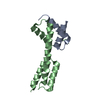
| ||||||||||||||||||||||||||||||||||||||||||||||||||||||||||||||||||||||||||||||||||||||||||||||||||||||||||||||||||||||||||||||||||||||||||||||||||||||||||||||||||||||||||||||||
| 2 | 
| ||||||||||||||||||||||||||||||||||||||||||||||||||||||||||||||||||||||||||||||||||||||||||||||||||||||||||||||||||||||||||||||||||||||||||||||||||||||||||||||||||||||||||||||||
| Unit cell |
| ||||||||||||||||||||||||||||||||||||||||||||||||||||||||||||||||||||||||||||||||||||||||||||||||||||||||||||||||||||||||||||||||||||||||||||||||||||||||||||||||||||||||||||||||
| Noncrystallographic symmetry (NCS) | NCS domain:
NCS domain segments: Component-ID: _ / Refine code: _
NCS ensembles :
|
-Components
| #1: Antibody | Mass: 11130.393 Da / Num. of mol.: 4 / Fragment: UNP RESIDUES 5-44,UNP RESIDUES 217-269 / Mutation: G240A Source method: isolated from a genetically manipulated source Details: The fusion protein of 4 tags, PKA (UNP residues 5-44) and Protein A (UNP residues 217-269) Source: (gene. exp.) Homo sapiens (human), (gene. exp.) Staphylococcus aureus (bacteria)Gene: PRKAR2A, PKR2, PRKAR2, spa / Production host: |
|---|
-Experimental details
-Experiment
| Experiment | Method: X-RAY DIFFRACTION / Number of used crystals: 1 |
|---|
- Sample preparation
| Crystal | Density Matthews: 3.85 Å3/Da / Density % sol: 68.05 % |
|---|---|
| Crystal grow | Temperature: 296 K / Method: vapor diffusion / pH: 8.5 / Details: 100mM Tris pH 8.5, 2.16M Sodium formate |
-Data collection
| Diffraction | Mean temperature: 80 K |
|---|---|
| Diffraction source | Source: SYNCHROTRON / Site: PAL/PLS  / Beamline: 7A (6B, 6C1) / Wavelength: 0.97934 Å / Beamline: 7A (6B, 6C1) / Wavelength: 0.97934 Å |
| Detector | Type: ADSC QUANTUM 315r / Detector: CCD / Date: Mar 16, 2017 |
| Radiation | Protocol: SINGLE WAVELENGTH / Monochromatic (M) / Laue (L): M / Scattering type: x-ray |
| Radiation wavelength | Wavelength: 0.97934 Å / Relative weight: 1 |
| Reflection | Resolution: 3.25→50 Å / Num. obs: 10889 / % possible obs: 98.9 % / Redundancy: 6.3 % / Net I/σ(I): 23.6 |
| Reflection shell | Resolution: 3.3→3.42 Å |
- Processing
| Software |
| ||||||||||||||||||||||||||||||||||||||||||||||||||||||||||||||||||||||||||||||||||||||||||||||||||||||||||||||||||||||||||||||||||||||||||||||||||||||||||||||||||||||||||||||||||||||
|---|---|---|---|---|---|---|---|---|---|---|---|---|---|---|---|---|---|---|---|---|---|---|---|---|---|---|---|---|---|---|---|---|---|---|---|---|---|---|---|---|---|---|---|---|---|---|---|---|---|---|---|---|---|---|---|---|---|---|---|---|---|---|---|---|---|---|---|---|---|---|---|---|---|---|---|---|---|---|---|---|---|---|---|---|---|---|---|---|---|---|---|---|---|---|---|---|---|---|---|---|---|---|---|---|---|---|---|---|---|---|---|---|---|---|---|---|---|---|---|---|---|---|---|---|---|---|---|---|---|---|---|---|---|---|---|---|---|---|---|---|---|---|---|---|---|---|---|---|---|---|---|---|---|---|---|---|---|---|---|---|---|---|---|---|---|---|---|---|---|---|---|---|---|---|---|---|---|---|---|---|---|---|---|
| Refinement | Method to determine structure: MOLECULAR REPLACEMENT Starting model: 2IZX, 4ZNC Resolution: 3.25→43.537 Å / Cor.coef. Fo:Fc: 0.92 / Cor.coef. Fo:Fc free: 0.871 / SU B: 75.516 / SU ML: 0.533 / Cross valid method: THROUGHOUT / ESU R Free: 0.509 / Stereochemistry target values: MAXIMUM LIKELIHOOD
| ||||||||||||||||||||||||||||||||||||||||||||||||||||||||||||||||||||||||||||||||||||||||||||||||||||||||||||||||||||||||||||||||||||||||||||||||||||||||||||||||||||||||||||||||||||||
| Solvent computation | Ion probe radii: 0.8 Å / Shrinkage radii: 0.8 Å / VDW probe radii: 1.2 Å / Solvent model: MASK | ||||||||||||||||||||||||||||||||||||||||||||||||||||||||||||||||||||||||||||||||||||||||||||||||||||||||||||||||||||||||||||||||||||||||||||||||||||||||||||||||||||||||||||||||||||||
| Displacement parameters | Biso mean: 165.823 Å2
| ||||||||||||||||||||||||||||||||||||||||||||||||||||||||||||||||||||||||||||||||||||||||||||||||||||||||||||||||||||||||||||||||||||||||||||||||||||||||||||||||||||||||||||||||||||||
| Refinement step | Cycle: 1 / Resolution: 3.25→43.537 Å
| ||||||||||||||||||||||||||||||||||||||||||||||||||||||||||||||||||||||||||||||||||||||||||||||||||||||||||||||||||||||||||||||||||||||||||||||||||||||||||||||||||||||||||||||||||||||
| Refine LS restraints |
|