 Movie
Movie Controller

[English] 日本語
 Yorodumi
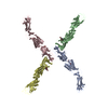
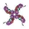
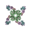
- PDB-5x3f: Crystal structure of the YgjG-Protein A-Zpa963-PKA catalytic domain -
+ Open data

- Basic information
| Entry | Database: PDB / ID: 5x3f | ||||||
|---|---|---|---|---|---|---|---|
| Title | Crystal structure of the YgjG-Protein A-Zpa963-PKA catalytic domain | ||||||
 Components Components |
| ||||||
 Keywords Keywords | TRANSFERASE / synthetic protein / LYASE | ||||||
| Function / homology |  Function and homology information Function and homology informationdiamine transaminase / putrescine-2-oxoglutarate transaminase / diamine:2-oxoglutarate transaminase activity / putrescine:2-oxoglutarate transaminase activity / putrescine catabolic process / L-lysine catabolic process / spontaneous exocytosis of neurotransmitter / PKA activation in glucagon signalling / CREB1 phosphorylation through the activation of Adenylate Cyclase / negative regulation of meiotic cell cycle ...diamine transaminase / putrescine-2-oxoglutarate transaminase / diamine:2-oxoglutarate transaminase activity / putrescine:2-oxoglutarate transaminase activity / putrescine catabolic process / L-lysine catabolic process / spontaneous exocytosis of neurotransmitter / PKA activation in glucagon signalling / CREB1 phosphorylation through the activation of Adenylate Cyclase / negative regulation of meiotic cell cycle / HDL assembly / positive regulation of triglyceride catabolic process / DARPP-32 events / Rap1 signalling / PKA activation / Regulation of insulin secretion / Vasopressin regulates renal water homeostasis via Aquaporins / GPER1 signaling / Glucagon-like Peptide-1 (GLP1) regulates insulin secretion / Hedgehog 'off' state / Loss of Nlp from mitotic centrosomes / Recruitment of mitotic centrosome proteins and complexes / Loss of proteins required for interphase microtubule organization from the centrosome / Anchoring of the basal body to the plasma membrane / Recruitment of NuMA to mitotic centrosomes / AURKA Activation by TPX2 / Factors involved in megakaryocyte development and platelet production / MAPK6/MAPK4 signaling / Interleukin-3, Interleukin-5 and GM-CSF signaling / CD209 (DC-SIGN) signaling / RET signaling / GLI3 is processed to GLI3R by the proteasome / High laminar flow shear stress activates signaling by PIEZO1 and PECAM1:CDH5:KDR in endothelial cells / Regulation of PLK1 Activity at G2/M Transition / Mitochondrial protein degradation / VEGFA-VEGFR2 Pathway / nucleotide-activated protein kinase complex / Ion homeostasis / regulation of cellular respiration / histone H1-4S35 kinase activity / cAMP-dependent protein kinase / IgG binding / regulation of protein processing / negative regulation of glycolytic process / cAMP-dependent protein kinase activity / cellular response to parathyroid hormone stimulus / protein localization to lipid droplet / regulation of bicellular tight junction assembly / cAMP-dependent protein kinase complex / interleukin-2-mediated signaling pathway / regulation of osteoblast differentiation / sperm capacitation / cellular response to cold / protein kinase A regulatory subunit binding / negative regulation of glycolytic process through fructose-6-phosphate / ciliary base / intracellular potassium ion homeostasis / mesoderm formation / TORC1 signaling / plasma membrane raft / cAMP/PKA signal transduction / axoneme / sperm flagellum / postsynaptic modulation of chemical synaptic transmission / regulation of synaptic transmission, glutamatergic / negative regulation of TORC1 signaling / protein serine/threonine/tyrosine kinase activity / regulation of proteasomal protein catabolic process / lipid droplet / cellular response to glucagon stimulus / cellular response to nutrient levels / positive regulation of phagocytosis / positive regulation of gluconeogenesis / acrosomal vesicle / positive regulation of protein export from nucleus / neuromuscular junction / negative regulation of smoothened signaling pathway / neural tube closure / cellular response to glucose stimulus / positive regulation of cholesterol biosynthetic process / modulation of chemical synaptic transmission / small GTPase binding / mRNA processing / adenylate cyclase-inhibiting G protein-coupled receptor signaling pathway / pyridoxal phosphate binding / manganese ion binding / cellular response to heat / adenylate cyclase-activating G protein-coupled receptor signaling pathway / presynapse / sperm midpiece / protein kinase activity / regulation of cell cycle / nuclear speck / postsynapse / protein domain specific binding / protein serine kinase activity / protein serine/threonine kinase activity / positive regulation of cell population proliferation / ubiquitin protein ligase binding / centrosome Similarity search - Function | ||||||
| Biological species |   Staphylococcus aureus (bacteria) Staphylococcus aureus (bacteria) | ||||||
| Method |  X-RAY DIFFRACTION / SYNCHROTRON / MOLECULAR REPLACEMENT / Resolution: 3.38 Å X-RAY DIFFRACTION / SYNCHROTRON / MOLECULAR REPLACEMENT / Resolution: 3.38 Å | ||||||
 Authors Authors | Youn, S.J. / Kwon, N.Y. / Lee, J.H. / Kim, J.H. / Lee, H. / Lee, J.O. | ||||||
 Citation Citation | Journal: Sci Rep / Year: 2017 Title: Construction of novel repeat proteins with rigid and predictable structures using a shared helix method. Authors: Youn, S.J. / Kwon, N.Y. / Lee, J.H. / Kim, J.H. / Choi, J. / Lee, H. / Lee, J.O. | ||||||
| History |
|
- Structure visualization
| Structure viewer | Molecule: MolmilJmol/JSmol |
|---|
- Downloads & links
-Download
| PDBx/mmCIF format | 5x3f.cif.gz | 352.1 KB | Display | PDBx/mmCIF format |
|---|---|---|---|---|
| PDB format | pdb5x3f.ent.gz | 290 KB | Display | PDB format |
| PDBx/mmJSON format | 5x3f.json.gz | Tree view | PDBx/mmJSON format | |
| Others |  Other downloads Other downloads |
-Validation report
| Arichive directory | https://data.pdbj.org/pub/pdb/validation_reports/x3/5x3fftp://data.pdbj.org/pub/pdb/validation_reports/x3/5x3f | HTTPS FTP |
|---|
-Related structure data
| Related structure data |  5h75C  5h76C 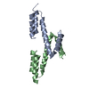 5h77C 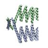 5h78C  5h79C  5h7aC  5h7bC  5h7cC  5h7dC  5xbyC  2m5aS  4ntsS 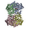 4uoyS S: Starting model for refinement C: citing same article ( |
|---|---|
| Similar structure data |


-Links
 PDBj
PDBj


- Assembly
| Deposited unit | 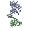
| ||||||||
|---|---|---|---|---|---|---|---|---|---|
| 1 | 
| ||||||||
| Unit cell |
|
-Components
| #1: Protein | Mass: 54451.414 Da / Num. of mol.: 1 / Fragment: UNP RESIDUES 7-453,220-269 / Mutation: N222V, G240A Source method: isolated from a genetically manipulated source Details: The fusion protein of Putrescine aminotransferase (UNP RESIDUES 7-453) and Immunoglobulin G-binding protein A (UNP RESIDUES 220-269) Source: (gene. exp.) Staphylococcus aureus (bacteria)Strain: K12 / Gene: patA, ygjG, b3073, JW5510, spa / Production host: References: UniProt: P42588, UniProt: P38507, putrescine-2-oxoglutarate transaminase |
|---|---|
| #2: Protein | Mass: 45479.789 Da / Num. of mol.: 1 / Fragment: residues 97-156,UNP RESIDUES 11-343 Source method: isolated from a genetically manipulated source Details: The fusion protein of Zpa963 and cAMP-dependent protein kinase catalytic subunit alpha (UNP RESIDUES 11-343) Source: (gene. exp.) Staphylococcus aureus (bacteria), (gene. exp.) Gene: Prkaca, Pkaca / Production host: |
| Has protein modification | Y |
-Experimental details
-Experiment
| Experiment | Method: X-RAY DIFFRACTION / Number of used crystals: 1 |
|---|
- Sample preparation
| Crystal | Density Matthews: 5.55 Å3/Da / Density % sol: 77.84 % |
|---|---|
| Crystal grow | Temperature: 296 K / Method: vapor diffusion / pH: 5.5 / Details: 91mM MES pH 5.5, 2.33M Na formate |
-Data collection
| Diffraction | Mean temperature: 80 K |
|---|---|
| Diffraction source | Source: SYNCHROTRON / Site: PAL/PLS  / Beamline: 7A (6B, 6C1) / Wavelength: 0.97934 Å / Beamline: 7A (6B, 6C1) / Wavelength: 0.97934 Å |
| Detector | Type: ADSC QUANTUM 315 / Detector: CCD / Date: Dec 14, 2016 |
| Radiation | Protocol: SINGLE WAVELENGTH / Monochromatic (M) / Laue (L): M / Scattering type: x-ray |
| Radiation wavelength | Wavelength: 0.97934 Å / Relative weight: 1 |
| Reflection | Resolution: 3.38→50 Å / Num. obs: 31056 / % possible obs: 99.7 % / Redundancy: 6.5 % / Net I/σ(I): 13.7 |
| Reflection shell | Resolution: 3.38→3.52 Å / Redundancy: 6.1 % / % possible all: 99.7 |
- Processing
| Software |
| ||||||||||||||||||||||||||||||||||||||||||||||||||||||||||||||||||||||||||||||||||||||||||||||||||||||||||||||||||||||||||||||||||||||||||||||||||||||||||||||||||||||||||||||||||||||
|---|---|---|---|---|---|---|---|---|---|---|---|---|---|---|---|---|---|---|---|---|---|---|---|---|---|---|---|---|---|---|---|---|---|---|---|---|---|---|---|---|---|---|---|---|---|---|---|---|---|---|---|---|---|---|---|---|---|---|---|---|---|---|---|---|---|---|---|---|---|---|---|---|---|---|---|---|---|---|---|---|---|---|---|---|---|---|---|---|---|---|---|---|---|---|---|---|---|---|---|---|---|---|---|---|---|---|---|---|---|---|---|---|---|---|---|---|---|---|---|---|---|---|---|---|---|---|---|---|---|---|---|---|---|---|---|---|---|---|---|---|---|---|---|---|---|---|---|---|---|---|---|---|---|---|---|---|---|---|---|---|---|---|---|---|---|---|---|---|---|---|---|---|---|---|---|---|---|---|---|---|---|---|---|
| Refinement | Method to determine structure: MOLECULAR REPLACEMENT Starting model: 4UOY, 2M5A, 4NTS Resolution: 3.38→50 Å / Cor.coef. Fo:Fc: 0.946 / Cor.coef. Fo:Fc free: 0.939 / SU B: 42.478 / SU ML: 0.288 / Cross valid method: THROUGHOUT / ESU R Free: 0.371 / Stereochemistry target values: MAXIMUM LIKELIHOOD
| ||||||||||||||||||||||||||||||||||||||||||||||||||||||||||||||||||||||||||||||||||||||||||||||||||||||||||||||||||||||||||||||||||||||||||||||||||||||||||||||||||||||||||||||||||||||
| Solvent computation | Ion probe radii: 0.8 Å / Shrinkage radii: 0.8 Å / VDW probe radii: 1.2 Å / Solvent model: MASK | ||||||||||||||||||||||||||||||||||||||||||||||||||||||||||||||||||||||||||||||||||||||||||||||||||||||||||||||||||||||||||||||||||||||||||||||||||||||||||||||||||||||||||||||||||||||
| Displacement parameters | Biso mean: 135.845 Å2
| ||||||||||||||||||||||||||||||||||||||||||||||||||||||||||||||||||||||||||||||||||||||||||||||||||||||||||||||||||||||||||||||||||||||||||||||||||||||||||||||||||||||||||||||||||||||
| Refinement step | Cycle: 1 / Resolution: 3.38→50 Å
| ||||||||||||||||||||||||||||||||||||||||||||||||||||||||||||||||||||||||||||||||||||||||||||||||||||||||||||||||||||||||||||||||||||||||||||||||||||||||||||||||||||||||||||||||||||||
| Refine LS restraints |
|