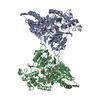
 Movie
Movie Controller


+ Open data

- Basic information
| Entry | Database: PDB / ID: 4uoy | ||||||
|---|---|---|---|---|---|---|---|
| Title | Crystal structure of YgjG in complex with Pyridoxal-5'-phosphate | ||||||
 Components Components | PUTRESCINE AMINOTRANSFERASE | ||||||
 Keywords Keywords | TRANSFERASE | ||||||
| Function / homology |  Function and homology information Function and homology informationdiamine transaminase / putrescine-2-oxoglutarate transaminase / diamine:2-oxoglutarate transaminase activity / putrescine:2-oxoglutarate transaminase activity / putrescine catabolic process / L-lysine catabolic process / pyridoxal phosphate binding / protein homodimerization activity / identical protein binding / cytosol Similarity search - Function | ||||||
| Biological species |  | ||||||
| Method |  X-RAY DIFFRACTION / SYNCHROTRON / MOLECULAR REPLACEMENT / Resolution: 2.305 Å X-RAY DIFFRACTION / SYNCHROTRON / MOLECULAR REPLACEMENT / Resolution: 2.305 Å | ||||||
 Authors Authors | Jeong, J.H. / Kim, Y.G. | ||||||
 Citation Citation | Journal: Plos One / Year: 2014 Title: Structure of Putrescine Aminotransferase from Escherichia Coli Provides Insights Into the Substrate Specificity Among Class III Aminotransferases. Authors: Cha, H.J. / Jeong, J. / Rojviriya, C. / Kim, Y. | ||||||
| History |
|
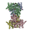
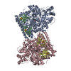
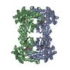
- Structure visualization
| Structure viewer | Molecule: MolmilJmol/JSmol |
|---|
- Downloads & links
-Download
| PDBx/mmCIF format | 4uoy.cif.gz | 345.5 KB | Display | PDBx/mmCIF format |
|---|---|---|---|---|
| PDB format | pdb4uoy.ent.gz | 282.7 KB | Display | PDB format |
| PDBx/mmJSON format | 4uoy.json.gz | Tree view | PDBx/mmJSON format | |
| Others |  Other downloads Other downloads |
-Validation report
| Arichive directory | https://data.pdbj.org/pub/pdb/validation_reports/uo/4uoyftp://data.pdbj.org/pub/pdb/validation_reports/uo/4uoy | HTTPS FTP |
|---|
-Related structure data
| Related structure data |  4uoxC 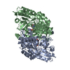 1vefS C: citing same article ( S: Starting model for refinement |
|---|---|
| Similar structure data |
-Links
 PDBj
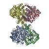
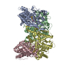
PDBj- Assembly
| Deposited unit | 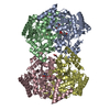
| ||||||||
|---|---|---|---|---|---|---|---|---|---|
| 1 |
| ||||||||
| Unit cell |
|
-Components
| #1: Protein | Mass: 50785.324 Da / Num. of mol.: 4 Source method: isolated from a genetically manipulated source Details: COVALENT BOND BETWEEN K 300 AND PYRIDOXAL-5'-PHOSPHATE Source: (gene. exp.) References: UniProt: P42588, putrescine-2-oxoglutarate transaminase #2: Chemical | ChemComp-PLP /   Mass: 247.142 Da / Num. of mol.: 4 / Source method: obtained synthetically / Formula: C8H10NO6P Mass: 247.142 Da / Num. of mol.: 4 / Source method: obtained synthetically / Formula: C8H10NO6P#3: Chemical | ChemComp-FMT / |   Mass: 46.025 Da / Num. of mol.: 1 / Source method: obtained synthetically / Formula: CH2O2 Mass: 46.025 Da / Num. of mol.: 1 / Source method: obtained synthetically / Formula: CH2O2#4: Chemical |   Mass: 92.094 Da / Num. of mol.: 2 / Source method: obtained synthetically / Formula: C3H8O3 Mass: 92.094 Da / Num. of mol.: 2 / Source method: obtained synthetically / Formula: C3H8O3#5: Water | ChemComp-HOH / |  Mass: 18.015 Da / Num. of mol.: 351 / Source method: isolated from a natural source / Formula: H2O Mass: 18.015 Da / Num. of mol.: 351 / Source method: isolated from a natural source / Formula: H2ONonpolymer details | PYRIDOXAL-5'-PHOSPHATE (PLP): PYRIDOXAL-5'-PHOSPHATE IS COVALENTLY | |
|---|
-Experimental details
-Experiment
| Experiment | Method: X-RAY DIFFRACTION / Number of used crystals: 1 |
|---|
- Sample preparation
| Crystal | Density Matthews: 2.58 Å3/Da / Density % sol: 52.44 % / Description: NONE |
|---|---|
| Crystal grow | pH: 7.5 Details: 0.1 M HEPES PH 7.5 15% PEG 3350 0.2 M SODIUM FORMATE 0.1 MM N-DODECYL-N,N-DIMETHYLGLYCINE |
-Data collection
| Diffraction | Mean temperature: 100 K |
|---|---|
| Diffraction source | Source: SYNCHROTRON / Site: PAL/PLS  / Beamline: 5C (4A) / Wavelength: 0.9795 / Beamline: 5C (4A) / Wavelength: 0.9795 |
| Detector | Type: ADSC QUANTUM 315 / Detector: CCD / Date: Nov 3, 2013 / Details: MIRRORS |
| Radiation | Monochromator: DOUBLE CRYSTAL MONOCHROMATOR / Protocol: SINGLE WAVELENGTH / Monochromatic (M) / Laue (L): M / Scattering type: x-ray |
| Radiation wavelength | Wavelength: 0.9795 Å / Relative weight: 1 |
| Reflection | Resolution: 2.3→30 Å / Num. obs: 91029 / % possible obs: 98.3 % / Observed criterion σ(I): 0 / Redundancy: 6.1 % / Biso Wilson estimate: 23.17 Å2 / Rmerge(I) obs: 0.1 / Net I/σ(I): 13.4 |
| Reflection shell | Resolution: 2.3→2.34 Å / Redundancy: 4.3 % / Rmerge(I) obs: 0.36 / Mean I/σ(I) obs: 3 / % possible all: 96.9 |
- Processing
| Software |
| |||||||||||||||||||||||||||||||||||||||||||||||||||||||||||||||||||||||||||||||||||||||||||||||||||||||||
|---|---|---|---|---|---|---|---|---|---|---|---|---|---|---|---|---|---|---|---|---|---|---|---|---|---|---|---|---|---|---|---|---|---|---|---|---|---|---|---|---|---|---|---|---|---|---|---|---|---|---|---|---|---|---|---|---|---|---|---|---|---|---|---|---|---|---|---|---|---|---|---|---|---|---|---|---|---|---|---|---|---|---|---|---|---|---|---|---|---|---|---|---|---|---|---|---|---|---|---|---|---|---|---|---|---|---|
| Refinement | Method to determine structure: MOLECULAR REPLACEMENT Starting model: PDB ENTRY 1VEF Resolution: 2.305→29.746 Å / SU ML: 0.34 / σ(F): 1.49 / Phase error: 21.45 / Stereochemistry target values: ML
| |||||||||||||||||||||||||||||||||||||||||||||||||||||||||||||||||||||||||||||||||||||||||||||||||||||||||
| Solvent computation | Shrinkage radii: 0.9 Å / VDW probe radii: 1.11 Å / Solvent model: FLAT BULK SOLVENT MODEL | |||||||||||||||||||||||||||||||||||||||||||||||||||||||||||||||||||||||||||||||||||||||||||||||||||||||||
| Refinement step | Cycle: LAST / Resolution: 2.305→29.746 Å
| |||||||||||||||||||||||||||||||||||||||||||||||||||||||||||||||||||||||||||||||||||||||||||||||||||||||||
| Refine LS restraints |
| |||||||||||||||||||||||||||||||||||||||||||||||||||||||||||||||||||||||||||||||||||||||||||||||||||||||||
| LS refinement shell |
|
