 Movie
Movie Controller

[English] 日本語
 Yorodumi
- PDB-5vbt: Crystal structure of a highly specific and potent USP7 ubiquitin ... -
+ Open data

- Basic information
| Entry | Database: PDB / ID: 5vbt | ||||||
|---|---|---|---|---|---|---|---|
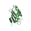
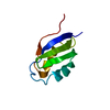
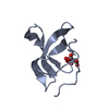
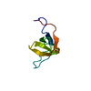
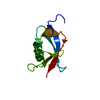
| Title | Crystal structure of a highly specific and potent USP7 ubiquitin variant inhibitor | ||||||
 Components Components | UBH04 | ||||||
 Keywords Keywords | DE NOVO PROTEIN / Ubiquitin / inhibitor / Structural Genomics / Structural Genomics Consortium / SGC | ||||||
| Biological species | synthetic construct (others) | ||||||
| Method |  X-RAY DIFFRACTION / SYNCHROTRON / MOLECULAR REPLACEMENT / Resolution: 1.51 Å X-RAY DIFFRACTION / SYNCHROTRON / MOLECULAR REPLACEMENT / Resolution: 1.51 Å | ||||||
 Authors Authors | DONG, A. / DONG, X. / LIU, L. / GUO, Y. / LI, Y. / ZHANG, W. / WALKER, J.R. / SIDHU, S. / Bountra, C. / Arrowsmith, C.H. ...DONG, A. / DONG, X. / LIU, L. / GUO, Y. / LI, Y. / ZHANG, W. / WALKER, J.R. / SIDHU, S. / Bountra, C. / Arrowsmith, C.H. / Edwards, A.M. / TONG, Y. / Structural Genomics Consortium (SGC) | ||||||
 Citation Citation | Journal: to be published Title: Crystal structure of a highly specific and potent USP7 ubiquitin variant inhibitor Authors: DONG, X. / DONG, A. / LIU, L. / GUO, Y. / LI, Y. / ZHANG, W. / WALKER, J.R. / SIDHU, S. / Bountra, C. / Arrowsmith, C.H. / Edwards, A.M. / TONG, Y. / Structural Genomics Consortium (SGC) | ||||||
| History |
|
- Structure visualization
| Structure viewer | Molecule:  MolmilJmol/JSmol MolmilJmol/JSmol |
|---|
- Downloads & links
-Download
| PDBx/mmCIF format | 5vbt.cif.gz | 48.9 KB | Display | PDBx/mmCIF format |
|---|---|---|---|---|
| PDB format | pdb5vbt.ent.gz | 33.1 KB | Display | PDB format |
| PDBx/mmJSON format | 5vbt.json.gz | Tree view | PDBx/mmJSON format | |
| Others |  Other downloads Other downloads |
-Validation report
| Arichive directory | https://data.pdbj.org/pub/pdb/validation_reports/vb/5vbtftp://data.pdbj.org/pub/pdb/validation_reports/vb/5vbt | HTTPS FTP |
|---|
-Related structure data
| Related structure data |  4ap4S S: Starting model for refinement |
|---|---|
| Similar structure data |


-Links
 PDBj
PDBj

- Assembly
| Deposited unit | 
| ||||||||
|---|---|---|---|---|---|---|---|---|---|
| 1 | 
| ||||||||
| 2 | 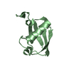
| ||||||||
| Unit cell |
|
-Components
| #1: Protein | Mass: 8876.288 Da / Num. of mol.: 2 Source method: isolated from a genetically manipulated source Source: (gene. exp.) synthetic construct (others) / Plasmid: pET28-MHL / Production host:  #2: Water | ChemComp-HOH / |  Mass: 18.015 Da / Num. of mol.: 156 / Source method: isolated from a natural source / Formula: H2O Mass: 18.015 Da / Num. of mol.: 156 / Source method: isolated from a natural source / Formula: H2O |
|---|
-Experimental details
-Experiment
| Experiment | Method: X-RAY DIFFRACTION / Number of used crystals: 1 |
|---|
- Sample preparation
| Crystal | Density Matthews: 2.02 Å3/Da / Density % sol: 39.2 % |
|---|---|
| Crystal grow | Temperature: 293 K / Method: vapor diffusion, sitting drop / pH: 8.5 / Details: 1.2 M sodium citrate, 0.1 M Tris pH 8.5 |
-Data collection
| Diffraction | Mean temperature: 100 K | |||||||||||||||||||||||||||||||||||||||||||||||||||||||||||||||||||||||||||||||||||||||||||||||||||||||||||||||||||||||||||||||||||||||||||||||||||||||||||||||||||||||||||||||||||||||||||||
|---|---|---|---|---|---|---|---|---|---|---|---|---|---|---|---|---|---|---|---|---|---|---|---|---|---|---|---|---|---|---|---|---|---|---|---|---|---|---|---|---|---|---|---|---|---|---|---|---|---|---|---|---|---|---|---|---|---|---|---|---|---|---|---|---|---|---|---|---|---|---|---|---|---|---|---|---|---|---|---|---|---|---|---|---|---|---|---|---|---|---|---|---|---|---|---|---|---|---|---|---|---|---|---|---|---|---|---|---|---|---|---|---|---|---|---|---|---|---|---|---|---|---|---|---|---|---|---|---|---|---|---|---|---|---|---|---|---|---|---|---|---|---|---|---|---|---|---|---|---|---|---|---|---|---|---|---|---|---|---|---|---|---|---|---|---|---|---|---|---|---|---|---|---|---|---|---|---|---|---|---|---|---|---|---|---|---|---|---|---|---|
| Diffraction source | Source: SYNCHROTRON / Site: APS  / Beamline: 19-ID / Wavelength: 0.97891 Å / Beamline: 19-ID / Wavelength: 0.97891 Å | |||||||||||||||||||||||||||||||||||||||||||||||||||||||||||||||||||||||||||||||||||||||||||||||||||||||||||||||||||||||||||||||||||||||||||||||||||||||||||||||||||||||||||||||||||||||||||||
| Detector | Type: DECTRIS PILATUS3 X 6M / Detector: PIXEL / Date: Dec 16, 2016 | |||||||||||||||||||||||||||||||||||||||||||||||||||||||||||||||||||||||||||||||||||||||||||||||||||||||||||||||||||||||||||||||||||||||||||||||||||||||||||||||||||||||||||||||||||||||||||||
| Radiation | Protocol: SINGLE WAVELENGTH / Monochromatic (M) / Laue (L): M / Scattering type: x-ray | |||||||||||||||||||||||||||||||||||||||||||||||||||||||||||||||||||||||||||||||||||||||||||||||||||||||||||||||||||||||||||||||||||||||||||||||||||||||||||||||||||||||||||||||||||||||||||||
| Radiation wavelength | Wavelength: 0.97891 Å / Relative weight: 1 | |||||||||||||||||||||||||||||||||||||||||||||||||||||||||||||||||||||||||||||||||||||||||||||||||||||||||||||||||||||||||||||||||||||||||||||||||||||||||||||||||||||||||||||||||||||||||||||
| Reflection | Resolution: 1.5→50 Å / Num. obs: 22616 / % possible obs: 98.8 % / Redundancy: 5.3 % / Rmerge(I) obs: 0.085 / Rpim(I) all: 0.04 / Rrim(I) all: 0.094 / Χ2: 0.899 / Net I/σ(I): 5.9 / Num. measured all: 119284 | |||||||||||||||||||||||||||||||||||||||||||||||||||||||||||||||||||||||||||||||||||||||||||||||||||||||||||||||||||||||||||||||||||||||||||||||||||||||||||||||||||||||||||||||||||||||||||||
| Reflection shell | Diffraction-ID: 1
|
- Processing
| Software |
| ||||||||||||||||||||||||||||||||||||||||||||||||||||||||||||
|---|---|---|---|---|---|---|---|---|---|---|---|---|---|---|---|---|---|---|---|---|---|---|---|---|---|---|---|---|---|---|---|---|---|---|---|---|---|---|---|---|---|---|---|---|---|---|---|---|---|---|---|---|---|---|---|---|---|---|---|---|---|
| Refinement | Method to determine structure: MOLECULAR REPLACEMENT Starting model: 4AP4 Resolution: 1.51→50.01 Å / Cor.coef. Fo:Fc: 0.971 / Cor.coef. Fo:Fc free: 0.959 / SU B: 1.455 / SU ML: 0.053 / Cross valid method: THROUGHOUT / σ(F): 0 / ESU R: 0.076 / ESU R Free: 0.077 / Stereochemistry target values: MAXIMUM LIKELIHOOD Details: HYDROGENS HAVE BEEN ADDED IN THE RIDING POSITIONS U VALUES : REFINED INDIVIDUALLY
| ||||||||||||||||||||||||||||||||||||||||||||||||||||||||||||
| Solvent computation | Ion probe radii: 0.8 Å / Shrinkage radii: 0.8 Å / VDW probe radii: 1.2 Å / Solvent model: MASK | ||||||||||||||||||||||||||||||||||||||||||||||||||||||||||||
| Displacement parameters | Biso max: 46.44 Å2 / Biso mean: 18.028 Å2 / Biso min: 11.68 Å2
| ||||||||||||||||||||||||||||||||||||||||||||||||||||||||||||
| Refinement step | Cycle: final / Resolution: 1.51→50.01 Å
| ||||||||||||||||||||||||||||||||||||||||||||||||||||||||||||
| Refine LS restraints |
| ||||||||||||||||||||||||||||||||||||||||||||||||||||||||||||
| LS refinement shell | Resolution: 1.505→1.544 Å / Rfactor Rfree error: 0 / Total num. of bins used: 20
|
