 Movie
Movie Controller


+ Open data

- Basic information
| Entry | Database: PDB / ID: 6exx | ||||||
|---|---|---|---|---|---|---|---|
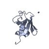
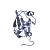
| Title | Crystal Structure of Pes4 RRM4 | ||||||
 Components Components | Protein PES4 | ||||||
 Keywords Keywords | RNA BINDING PROTEIN / RNA Recognition Motif | ||||||
| Function / homology |  Function and homology information Function and homology informationmRNA localization resulting in post-transcriptional regulation of gene expression / Deadenylation of mRNA / sporulation / prospore membrane / mRNA metabolic process / poly(A) binding / poly(U) RNA binding / Translation initiation complex formation / Nonsense Mediated Decay (NMD) independent of the Exon Junction Complex (EJC) / Nonsense Mediated Decay (NMD) enhanced by the Exon Junction Complex (EJC) ...mRNA localization resulting in post-transcriptional regulation of gene expression / Deadenylation of mRNA / sporulation / prospore membrane / mRNA metabolic process / poly(A) binding / poly(U) RNA binding / Translation initiation complex formation / Nonsense Mediated Decay (NMD) independent of the Exon Junction Complex (EJC) / Nonsense Mediated Decay (NMD) enhanced by the Exon Junction Complex (EJC) / L13a-mediated translational silencing of Ceruloplasmin expression / mRNA 3'-UTR binding / cytoplasmic stress granule / regulation of translation / ribonucleoprotein complex / nucleus / cytosol Similarity search - Function | ||||||
| Biological species |  | ||||||
| Method |  X-RAY DIFFRACTION / SYNCHROTRON / Resolution: 1.1 Å X-RAY DIFFRACTION / SYNCHROTRON / Resolution: 1.1 Å | ||||||
 Authors Authors | Mohamad, N. / Bravo, J. | ||||||
| Funding support |  Spain, 1items Spain, 1items
| ||||||
 Citation Citation | Journal: To Be Published Title: Crystal Structure of Pes4 RRM4 at 1.1 Angstroms resolution Authors: Mohamad, N. / Bravo, J. | ||||||
| History |
|
- Structure visualization
| Structure viewer | Molecule: MolmilJmol/JSmol |
|---|
- Downloads & links
-Download
| PDBx/mmCIF format | 6exx.cif.gz | 60.2 KB | Display | PDBx/mmCIF format |
|---|---|---|---|---|
| PDB format | pdb6exx.ent.gz | 43.8 KB | Display | PDB format |
| PDBx/mmJSON format | 6exx.json.gz | Tree view | PDBx/mmJSON format | |
| Others |  Other downloads Other downloads |
-Validation report
| Arichive directory | https://data.pdbj.org/pub/pdb/validation_reports/ex/6exxftp://data.pdbj.org/pub/pdb/validation_reports/ex/6exx | HTTPS FTP |
|---|
-Related structure data
| Similar structure data |
|---|


-Links
 PDBj
PDBj


- Assembly
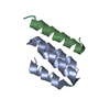
| Deposited unit | 
| ||||||||
|---|---|---|---|---|---|---|---|---|---|
| 1 |
| ||||||||
| Unit cell |
|
-Components
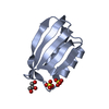
| #1: Protein | Mass: 9243.413 Da / Num. of mol.: 1 Source method: isolated from a genetically manipulated source Source: (gene. exp.) Strain: ATCC 204508 / S288c / Gene: PES4, YFR023W / Production host:  |
|---|---|
| #2: Water | ChemComp-HOH /  Mass: 18.015 Da / Num. of mol.: 104 / Source method: isolated from a natural source / Formula: H2O Mass: 18.015 Da / Num. of mol.: 104 / Source method: isolated from a natural source / Formula: H2O |
-Experimental details
-Experiment
| Experiment | Method: X-RAY DIFFRACTION / Number of used crystals: 1 |
|---|
- Sample preparation
| Crystal | Density Matthews: 1.98 Å3/Da / Density % sol: 37.85 % |
|---|---|
| Crystal grow | Temperature: 294 K / Method: vapor diffusion, sitting drop Details: 0.2 M Ammonium Nitrate 20% PEG 3350 0.1 M Betaine Monohydrate (as an additive) |
-Data collection
| Diffraction | Mean temperature: 100 K |
|---|---|
| Diffraction source | Source: SYNCHROTRON / Site: ALBA / Beamline: XALOC / Wavelength: 0.97921 Å |
| Detector | Type: DECTRIS PILATUS 6M / Detector: PIXEL / Date: Apr 15, 2016 |
| Radiation | Protocol: SINGLE WAVELENGTH / Monochromatic (M) / Laue (L): M / Scattering type: x-ray |
| Radiation wavelength | Wavelength: 0.97921 Å / Relative weight: 1 |
| Reflection | Resolution: 1.1→25.79 Å / Num. obs: 30587 / % possible obs: 99.8 % / Redundancy: 12.3 % / Rmerge(I) obs: 0.05842 / Rrim(I) all: 0.06103 / Net I/σ(I): 21.51 |
| Reflection shell | Resolution: 1.1→1.12 Å / Rmerge(I) obs: 0.6737 / % possible all: 100 |
- Processing
| Software |
| |||||||||||||||||||||||||||||||||||||||||||||||||||||||||||||||||||||||||||||
|---|---|---|---|---|---|---|---|---|---|---|---|---|---|---|---|---|---|---|---|---|---|---|---|---|---|---|---|---|---|---|---|---|---|---|---|---|---|---|---|---|---|---|---|---|---|---|---|---|---|---|---|---|---|---|---|---|---|---|---|---|---|---|---|---|---|---|---|---|---|---|---|---|---|---|---|---|---|---|
| Refinement | Resolution: 1.1→25.79 Å / SU ML: 0.08 / Cross valid method: FREE R-VALUE / σ(F): 1.34 / Phase error: 14.79
| |||||||||||||||||||||||||||||||||||||||||||||||||||||||||||||||||||||||||||||
| Solvent computation | Shrinkage radii: 0.9 Å / VDW probe radii: 1.11 Å | |||||||||||||||||||||||||||||||||||||||||||||||||||||||||||||||||||||||||||||
| Refinement step | Cycle: LAST / Resolution: 1.1→25.79 Å
| |||||||||||||||||||||||||||||||||||||||||||||||||||||||||||||||||||||||||||||
| Refine LS restraints |
| |||||||||||||||||||||||||||||||||||||||||||||||||||||||||||||||||||||||||||||
| LS refinement shell |
|
