Movie
Movie Controller

[English] 日本語
 Yorodumi
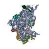
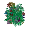
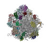
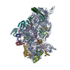
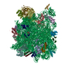
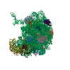
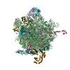
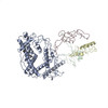
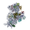
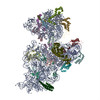
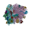
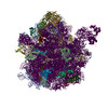
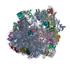
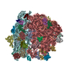
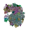
- PDB-4v48: Real space refined coordinates of the 30S and 50S subunits fitted... -
+ Open data

- Basic information
| Entry | Database: PDB / ID: 4v48 | |||||||||
|---|---|---|---|---|---|---|---|---|---|---|
| Title | Real space refined coordinates of the 30S and 50S subunits fitted into the low resolution cryo-EM map of the initiation-like state of E. coli 70S ribosome | |||||||||
 Components Components |
| |||||||||
 Keywords Keywords | RIBOSOME / EF-G.GTP-bound state / ratchet-like movement / real-space refinement | |||||||||
| Function / homology |  Function and homology information Function and homology informationstringent response / translational termination / transcriptional attenuation / endoribonuclease inhibitor activity / positive regulation of ribosome biogenesis / RNA-binding transcription regulator activity / four-way junction DNA binding / negative regulation of cytoplasmic translation / DnaA-L2 complex / translation repressor activity ...stringent response / translational termination / transcriptional attenuation / endoribonuclease inhibitor activity / positive regulation of ribosome biogenesis / RNA-binding transcription regulator activity / four-way junction DNA binding / negative regulation of cytoplasmic translation / DnaA-L2 complex / translation repressor activity / negative regulation of DNA-templated DNA replication initiation / mRNA regulatory element binding translation repressor activity / response to reactive oxygen species / cytosolic ribosome assembly / ribosome assembly / assembly of large subunit precursor of preribosome / maturation of SSU-rRNA from tricistronic rRNA transcript (SSU-rRNA, 5.8S rRNA, LSU-rRNA) / DNA endonuclease activity / regulation of cell growth / DNA-templated transcription termination / response to radiation / mRNA 5'-UTR binding / regulation of translation / large ribosomal subunit / transferase activity / ribosomal small subunit assembly / ribosome binding / ribosomal small subunit biogenesis / 5S rRNA binding / ribosomal large subunit assembly / small ribosomal subunit / small ribosomal subunit rRNA binding / cytosolic small ribosomal subunit / large ribosomal subunit rRNA binding / cytosolic large ribosomal subunit / cytoplasmic translation / tRNA binding / negative regulation of translation / rRNA binding / structural constituent of ribosome / ribosome / translation / ribonucleoprotein complex / response to antibiotic / negative regulation of DNA-templated transcription / hydrolase activity / mRNA binding / DNA binding / RNA binding / zinc ion binding / membrane / cytosol / cytoplasm Similarity search - Function | |||||||||
| Biological species |  | |||||||||
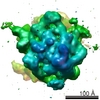
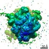
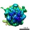
| Method | ELECTRON MICROSCOPY / single particle reconstruction / cryo EM / Resolution: 11.5 Å | |||||||||
 Authors Authors | Gao, H. / Sengupta, J. / Valle, M. / Korostelev, A. / Eswar, N. / Stagg, S.M. / Van Roey, P. / Agrawal, R.K. / Harvey, S.T. / Sali, A. ...Gao, H. / Sengupta, J. / Valle, M. / Korostelev, A. / Eswar, N. / Stagg, S.M. / Van Roey, P. / Agrawal, R.K. / Harvey, S.T. / Sali, A. / Chapman, M.S. / Frank, J. | |||||||||
 Citation Citation | Journal: Cell / Year: 2003 Title: Study of the structural dynamics of the E coli 70S ribosome using real-space refinement. Authors: Haixiao Gao / Jayati Sengupta / Mikel Valle / Andrei Korostelev / Narayanan Eswar / Scott M Stagg / Patrick Van Roey / Rajendra K Agrawal / Stephen C Harvey / Andrej Sali / Michael S Chapman / Joachim Frank /  Abstract: Cryo-EM density maps showing the 70S ribosome of E. coli in two different functional states related by a ratchet-like motion were analyzed using real-space refinement. Comparison of the two resulting ...Cryo-EM density maps showing the 70S ribosome of E. coli in two different functional states related by a ratchet-like motion were analyzed using real-space refinement. Comparison of the two resulting atomic models shows that the ribosome changes from a compact structure to a looser one, coupled with the rearrangement of many of the proteins. Furthermore, in contrast to the unchanged inter-subunit bridges formed wholly by RNA, the bridges involving proteins undergo large conformational changes following the ratchet-like motion, suggesting an important role of ribosomal proteins in facilitating the dynamics of translation. #1: Journal: Cell(Cambridge,Mass.) / Year: 2000Title: Solution structure of the E. coli 70S ribosome at 11.5 A resolution Authors: Gabashvili, I.S. / Agrawal, R.K. / Spahn, C.M.T. / Grassucci, R. / Svergun, D.I. / Frank, J. / Penczek, P. #2: Journal: Nature / Year: 2000Title: A ratchet-like inter-subunit reorganization of the ribosome during translocation Authors: Frank, J. / Agrawal, R.K. | |||||||||
| History |
|
- Structure visualization
| Movie |
Movie viewer |
|---|---|
| Structure viewer | Molecule: MolmilJmol/JSmol |
- Downloads & links
-Download
| PDBx/mmCIF format | 4v48.cif.gz | 332.4 KB | Display | PDBx/mmCIF format |
|---|---|---|---|---|
| PDB format | pdb4v48.ent.gz | Display | PDB format | |
| PDBx/mmJSON format | 4v48.json.gz | Tree view | PDBx/mmJSON format | |
| Others |  Other downloads Other downloads |
-Validation report
| Arichive directory | https://data.pdbj.org/pub/pdb/validation_reports/v4/4v48ftp://data.pdbj.org/pub/pdb/validation_reports/v4/4v48 | HTTPS FTP |
|---|
-Related structure data


-Links
 PDBj
PDBj


- Assembly
| Deposited unit | 
|
|---|---|
| 1 |
|
-Components
-RNA chain , 4 types, 4 molecules A0A9A6BA
| #1: RNA chain | Mass: 941612.375 Da / Num. of mol.: 1 / Source method: isolated from a natural source / Source: (natural) |
|---|---|
| #2: RNA chain | Mass: 38790.090 Da / Num. of mol.: 1 / Source method: isolated from a natural source / Source: (natural) |
| #3: RNA chain | Mass: 24518.570 Da / Num. of mol.: 1 / Source method: isolated from a natural source / Source: (natural) |
| #29: RNA chain | Mass: 499995.156 Da / Num. of mol.: 1 / Source method: isolated from a natural source / Source: (natural) |
+50S ribosomal protein ... , 25 types, 25 molecules AAABACADAEAFAGAHAIAJAKALAMANAOAQARASATAUAWAXAZA1A4
-30S RIBOSOMAL PROTEIN ... , 19 types, 19 molecules BBBCBDBEBFBGBHBIBJBKBLBMBNBOBPBQBRBSBT
| #30: Protein | Mass: 26650.475 Da / Num. of mol.: 1 / Source method: isolated from a natural source / Source: (natural) |
|---|---|
| #31: Protein | Mass: 25900.117 Da / Num. of mol.: 1 / Source method: isolated from a natural source / Source: (natural) |
| #32: Protein | Mass: 23383.002 Da / Num. of mol.: 1 / Source method: isolated from a natural source / Source: (natural) |
| #33: Protein | Mass: 17498.203 Da / Num. of mol.: 1 / Source method: isolated from a natural source / Source: (natural) |
| #34: Protein | Mass: 15727.512 Da / Num. of mol.: 1 / Source method: isolated from a natural source / Source: (natural) |
| #35: Protein | Mass: 19923.959 Da / Num. of mol.: 1 / Source method: isolated from a natural source / Source: (natural) |
| #36: Protein | Mass: 14015.361 Da / Num. of mol.: 1 / Source method: isolated from a natural source / Source: (natural) |
| #37: Protein | Mass: 14755.074 Da / Num. of mol.: 1 / Source method: isolated from a natural source / Source: (natural) |
| #38: Protein | Mass: 11755.597 Da / Num. of mol.: 1 / Source method: isolated from a natural source / Source: (natural) |
| #39: Protein | Mass: 13739.778 Da / Num. of mol.: 1 / Source method: isolated from a natural source / Source: (natural) |
| #40: Protein | Mass: 13636.961 Da / Num. of mol.: 1 / Source method: isolated from a natural source / Source: (natural) |
| #41: Protein | Mass: 12997.271 Da / Num. of mol.: 1 / Source method: isolated from a natural source / Source: (natural) |
| #42: Protein | Mass: 11475.364 Da / Num. of mol.: 1 / Source method: isolated from a natural source / Source: (natural) |
| #43: Protein | Mass: 10159.621 Da / Num. of mol.: 1 / Source method: isolated from a natural source / Source: (natural) |
| #44: Protein | Mass: 9207.572 Da / Num. of mol.: 1 / Source method: isolated from a natural source / Source: (natural) |
| #45: Protein | Mass: 9593.296 Da / Num. of mol.: 1 / Source method: isolated from a natural source / Source: (natural) |
| #46: Protein | Mass: 8874.276 Da / Num. of mol.: 1 / Source method: isolated from a natural source / Source: (natural) |
| #47: Protein | Mass: 10324.160 Da / Num. of mol.: 1 / Source method: isolated from a natural source / Source: (natural) |
| #48: Protein | Mass: 9577.268 Da / Num. of mol.: 1 / Source method: isolated from a natural source / Source: (natural) |
-Experimental details
-Experiment
| Experiment | Method: ELECTRON MICROSCOPY |
|---|---|
| EM experiment | Aggregation state: PARTICLE / 3D reconstruction method: single particle reconstruction |
- Sample preparation
| Component | Name: E. coli 70S ribosome bound with formyl-methionyl initiator tRNA Type: RIBOSOME |
|---|---|
| Buffer solution | Name: hepes / pH: 7.5 / Details: hepes |
| Specimen | Conc.: 32 mg/ml / Embedding applied: NO / Shadowing applied: NO / Staining applied: NO / Vitrification applied: YES |
| Vitrification | Details: Rapid-freezing in liquid ethane |
- Electron microscopy imaging
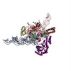
| Experimental equipment |  Model: Tecnai F20 / Image courtesy: FEI Company |
|---|---|
| Microscopy | Model: FEI TECNAI F20 / Date: Jul 1, 1997 |
| Electron gun | Electron source:  FIELD EMISSION GUN / Accelerating voltage: 200 kV / Illumination mode: FLOOD BEAM FIELD EMISSION GUN / Accelerating voltage: 200 kV / Illumination mode: FLOOD BEAM |
| Electron lens | Mode: BRIGHT FIELD / Nominal magnification: 51200 X / Calibrated magnification: 51200 X / Nominal defocus max: 4340 nm / Nominal defocus min: 730 nm / Cs: 2 mm |
| Specimen holder | Temperature: 93 K / Tilt angle max: 0 ° / Tilt angle min: 0 ° |
| Image recording | Electron dose: 10 e/Å2 / Film or detector model: KODAK SO-163 FILM |
- Processing
| EM software |
| |||||||||||||||||||||||||||||||||||||||||||||||||
|---|---|---|---|---|---|---|---|---|---|---|---|---|---|---|---|---|---|---|---|---|---|---|---|---|---|---|---|---|---|---|---|---|---|---|---|---|---|---|---|---|---|---|---|---|---|---|---|---|---|---|
| CTF correction | Details: CTF correction of 3D-maps | |||||||||||||||||||||||||||||||||||||||||||||||||
| Symmetry | Point symmetry: C1 (asymmetric) | |||||||||||||||||||||||||||||||||||||||||||||||||
| 3D reconstruction | Method: Reference based alignment / Resolution: 11.5 Å / Nominal pixel size: 2.93 Å / Actual pixel size: 2.93 Å / Magnification calibration: TMV / Details: SPIDER package / Symmetry type: POINT | |||||||||||||||||||||||||||||||||||||||||||||||||
| Atomic model building | Protocol: RIGID BODY FIT / Space: REAL Target criteria: cross-correlation coefficient, real space R factor Details: METHOD--auto REFINEMENT PROTOCOL--Multi-rigid body, real-space refinement | |||||||||||||||||||||||||||||||||||||||||||||||||
| Atomic model building |
| |||||||||||||||||||||||||||||||||||||||||||||||||
| Refinement step | Cycle: LAST
|