 Movie
Movie Controller

[English] 日本語
 Yorodumi
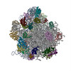
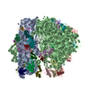
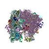
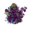
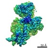
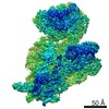
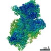
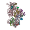
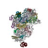
- PDB-4adv: Structure of the E. coli methyltransferase KsgA bound to the E. c... -
+ Open data

- Basic information
| Entry | Database: PDB / ID: 4adv | ||||||
|---|---|---|---|---|---|---|---|
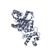
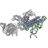
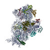
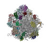
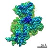
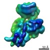
| Title | Structure of the E. coli methyltransferase KsgA bound to the E. coli 30S ribosomal subunit | ||||||
 Components Components |
| ||||||
 Keywords Keywords | TRANSLATION / RIBOSOME BIOGENESIS / SMALL RIBOSOMAL SUBUNIT | ||||||
| Function / homology |  Function and homology information Function and homology information16S rRNA (adenine1518-N6/adenine1519-N6)-dimethyltransferase / 16S rRNA (adenine(1518)-N(6)/adenine(1519)-N(6))-dimethyltransferase activity / rRNA (adenine-N6,N6-)-dimethyltransferase activity / rRNA base methylation / rRNA methylation / transcription antitermination factor activity, RNA binding / ornithine decarboxylase inhibitor activity / ribosomal small subunit binding / misfolded RNA binding / Group I intron splicing ...16S rRNA (adenine1518-N6/adenine1519-N6)-dimethyltransferase / 16S rRNA (adenine(1518)-N(6)/adenine(1519)-N(6))-dimethyltransferase activity / rRNA (adenine-N6,N6-)-dimethyltransferase activity / rRNA base methylation / rRNA methylation / transcription antitermination factor activity, RNA binding / ornithine decarboxylase inhibitor activity / ribosomal small subunit binding / misfolded RNA binding / Group I intron splicing / RNA folding / four-way junction DNA binding / regulation of mRNA stability / negative regulation of translational initiation / mRNA regulatory element binding translation repressor activity / positive regulation of RNA splicing / regulation of DNA-templated transcription elongation / transcription elongation factor complex / transcription antitermination / maturation of SSU-rRNA / DNA endonuclease activity / DNA-templated transcription termination / maintenance of translational fidelity / mRNA 5'-UTR binding / rRNA processing / regulation of translation / ribosomal small subunit assembly / ribosome biogenesis / ribosomal small subunit biogenesis / double-stranded DNA binding / small ribosomal subunit / small ribosomal subunit rRNA binding / cytosolic small ribosomal subunit / cytoplasmic translation / tRNA binding / negative regulation of translation / rRNA binding / structural constituent of ribosome / ribosome / translation / response to antibiotic / hydrolase activity / mRNA binding / RNA binding / zinc ion binding / membrane / cytoplasm / cytosol Similarity search - Function | ||||||
| Biological species |  | ||||||
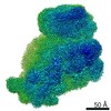
| Method | ELECTRON MICROSCOPY / single particle reconstruction / cryo EM / Resolution: 13.5 Å | ||||||
 Authors Authors | Boehringer, D. / O'Farrell, H.C. / Rife, J.P. / Ban, N. | ||||||
 Citation Citation | Journal: J Biol Chem / Year: 2012 Title: Structural insights into methyltransferase KsgA function in 30S ribosomal subunit biogenesis. Authors: Daniel Boehringer / Heather C O'Farrell / Jason P Rife / Nenad Ban /   Abstract: The assembly of the ribosomal subunits is facilitated by ribosome biogenesis factors. The universally conserved methyltransferase KsgA modifies two adjacent adenosine residues in the 3'-terminal ...The assembly of the ribosomal subunits is facilitated by ribosome biogenesis factors. The universally conserved methyltransferase KsgA modifies two adjacent adenosine residues in the 3'-terminal helix 45 of the 16 S ribosomal RNA (rRNA). KsgA recognizes its substrate adenosine residues only in the context of a near mature 30S subunit and is required for the efficient processing of the rRNA termini during ribosome biogenesis. Here, we present the cryo-EM structure of KsgA bound to a nonmethylated 30S ribosomal subunit. The structure reveals that KsgA binds to the 30S platform with the catalytic N-terminal domain interacting with substrate adenosine residues in helix 45 and the C-terminal domain making extensive contacts to helix 27 and helix 24. KsgA excludes the penultimate rRNA helix 44 from adopting its position in the mature 30S subunit, blocking the formation of the decoding site and subunit joining. We suggest that the activation of methyltransferase activity and subsequent dissociation of KsgA control conformational changes in helix 44 required for final rRNA processing and translation initiation. | ||||||
| History |
|
- Structure visualization
| Movie |
Movie viewer |
|---|---|
| Structure viewer | Molecule: MolmilJmol/JSmol |
- Downloads & links
-Download
| PDBx/mmCIF format | 4adv.cif.gz | 1.1 MB | Display | PDBx/mmCIF format |
|---|---|---|---|---|
| PDB format | pdb4adv.ent.gz | 901.2 KB | Display | PDB format |
| PDBx/mmJSON format | 4adv.json.gz | Tree view | PDBx/mmJSON format | |
| Others |  Other downloads Other downloads |
-Validation report
| Arichive directory | https://data.pdbj.org/pub/pdb/validation_reports/ad/4advftp://data.pdbj.org/pub/pdb/validation_reports/ad/4adv | HTTPS FTP |
|---|
-Related structure data
| Related structure data | 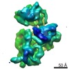 2017MC 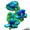 2019C 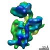 2020C M: map data used to model this data C: citing same article ( |
|---|---|
| Similar structure data |


-Links
 PDBj
PDBj


- Assembly
| Deposited unit | 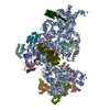
|
|---|---|
| 1 |
|
-Components
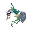
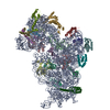
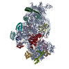
-30S RIBOSOMAL PROTEIN ... , 20 types, 20 molecules BCDEFGHIJKLMNOPQRSTU
| #2: Protein | Mass: 26650.475 Da / Num. of mol.: 1 / Source method: isolated from a natural source / Source: (natural) |
|---|---|
| #3: Protein | Mass: 25900.117 Da / Num. of mol.: 1 / Source method: isolated from a natural source / Source: (natural) |
| #4: Protein | Mass: 23383.002 Da / Num. of mol.: 1 / Source method: isolated from a natural source / Source: (natural) |
| #5: Protein | Mass: 17498.203 Da / Num. of mol.: 1 / Source method: isolated from a natural source / Source: (natural) |
| #6: Protein | Mass: 15727.512 Da / Num. of mol.: 1 / Source method: isolated from a natural source / Source: (natural) |
| #7: Protein | Mass: 19923.959 Da / Num. of mol.: 1 / Source method: isolated from a natural source / Source: (natural) |
| #8: Protein | Mass: 14015.361 Da / Num. of mol.: 1 / Source method: isolated from a natural source / Source: (natural) |
| #9: Protein | Mass: 14755.074 Da / Num. of mol.: 1 / Source method: isolated from a natural source / Source: (natural) |
| #10: Protein | Mass: 11755.597 Da / Num. of mol.: 1 / Source method: isolated from a natural source / Source: (natural) |
| #11: Protein | Mass: 13739.778 Da / Num. of mol.: 1 / Source method: isolated from a natural source / Source: (natural) |
| #12: Protein | Mass: 13636.961 Da / Num. of mol.: 1 / Source method: isolated from a natural source / Source: (natural) |
| #13: Protein | Mass: 12997.271 Da / Num. of mol.: 1 / Source method: isolated from a natural source / Source: (natural) |
| #14: Protein | Mass: 11475.364 Da / Num. of mol.: 1 / Source method: isolated from a natural source / Source: (natural) |
| #15: Protein | Mass: 10319.882 Da / Num. of mol.: 1 / Source method: isolated from a natural source / Source: (natural) |
| #16: Protein | Mass: 9207.572 Da / Num. of mol.: 1 / Source method: isolated from a natural source / Source: (natural) |
| #17: Protein | Mass: 9593.296 Da / Num. of mol.: 1 / Source method: isolated from a natural source / Source: (natural) |
| #18: Protein | Mass: 8874.276 Da / Num. of mol.: 1 / Source method: isolated from a natural source / Source: (natural) |
| #19: Protein | Mass: 10324.160 Da / Num. of mol.: 1 / Source method: isolated from a natural source / Source: (natural) |
| #20: Protein | Mass: 9577.268 Da / Num. of mol.: 1 / Source method: isolated from a natural source / Source: (natural) |
| #21: Protein | Mass: 8524.039 Da / Num. of mol.: 1 / Source method: isolated from a natural source / Source: (natural) |
-RNA chain / Protein / Non-polymers , 3 types, 141 molecules AV
| #1: RNA chain | Mass: 499690.031 Da / Num. of mol.: 1 / Source method: isolated from a natural source / Source: (natural) |
|---|---|
| #22: Protein | Mass: 27988.273 Da / Num. of mol.: 1 / Fragment: RESIDUES 17-268 Source method: isolated from a genetically manipulated source Source: (gene. exp.) References: UniProt: P06992, 16S rRNA (adenine1518-N6/adenine1519-N6)-dimethyltransferase |
| #23: Water | ChemComp-HOH / Mass: 18.015 Da / Num. of mol.: 139 / Source method: isolated from a natural source / Formula: H2O |
-Experimental details
-Experiment
| Experiment | Method: ELECTRON MICROSCOPY |
|---|---|
| EM experiment | Aggregation state: PARTICLE / 3D reconstruction method: single particle reconstruction |
- Sample preparation
| Component | Name: E. COLI METHYLTRANSFERASE KSGA BOUND TO THE E. COLI 30S RIBOSOMAL SUBUNIT Type: COMPLEX |
|---|---|
| Buffer solution | Name: 40 MM KCL, 4MM MGCL2, 20 MM HEPES/KOH PH 7. 6, 6 MM 2- MERCAPTOETHANOL pH: 7.6 Details: 40 MM KCL, 4MM MGCL2, 20 MM HEPES/KOH PH 7. 6, 6 MM 2- MERCAPTOETHANOL |
| Specimen | Embedding applied: NO / Shadowing applied: NO / Staining applied: NO / Vitrification applied: YES |
| Specimen support | Details: HOLEY CARBON |
| Vitrification | Instrument: HOMEMADE PLUNGER / Cryogen name: ETHANE Details: VITRIFICATION 1 -- CRYOGEN- ETHANE, INSTRUMENT- PLUNGER, |
- Electron microscopy imaging
| Microscopy | Model: FEI TECNAI 20 Details: SEMI-AUTOMATIC DATA ACQUISITION WITH SERIAL EM SCRIPT |
|---|---|
| Electron gun | Electron source:  FIELD EMISSION GUN / Accelerating voltage: 200 kV / Illumination mode: SPOT SCAN FIELD EMISSION GUN / Accelerating voltage: 200 kV / Illumination mode: SPOT SCAN |
| Electron lens | Mode: BRIGHT FIELD / Nominal magnification: 62000 X / Calibrated magnification: 82000 X / Nominal defocus max: 4000 nm / Nominal defocus min: 2000 nm |
| Image recording | Electron dose: 20 e/Å2 / Film or detector model: GATAN ULTRASCAN 4000 (4k x 4k) |
| Radiation wavelength | Relative weight: 1 |
- Processing
| EM software |
| |||||||||||||||||||||
|---|---|---|---|---|---|---|---|---|---|---|---|---|---|---|---|---|---|---|---|---|---|---|
| CTF correction | Details: PER IMAGE, CTFFIND3 | |||||||||||||||||||||
| Symmetry | Point symmetry: C1 (asymmetric) | |||||||||||||||||||||
| 3D reconstruction | Method: ANGULAR RECONSTITUTION, PROJECTION MATCHING / Resolution: 13.5 Å / Num. of particles: 23343 / Actual pixel size: 3.07 Å Details: 30S RIBOSOMAL SUBUNIT HEAD AND BODY WERE FITTED SEPARATELY AS RIGID BODIES. SUBMISSION BASED ON EXPERIMENTAL DATA FROM EMDB EMD-2017. (DEPOSITION ID: 10519). Symmetry type: POINT | |||||||||||||||||||||
| Atomic model building | Protocol: RIGID BODY FIT / Space: REAL / Target criteria: Cross-correlation coefficient / Details: METHOD--RIGID BODY REFINEMENT PROTOCOL--XRAY | |||||||||||||||||||||
| Atomic model building |
| |||||||||||||||||||||
| Refinement | Highest resolution: 13.5 Å | |||||||||||||||||||||
| Refinement step | Cycle: LAST / Highest resolution: 13.5 Å
|

