Monochromator: Osmic Confocal Optics / Protocol: SINGLE WAVELENGTH / Monochromatic (M) / Laue (L): M / Scattering type: x-ray
Radiation wavelength
Wavelength: 1.5418 Å / Relative weight: 1
Reflection
Resolution: 2.1→40 Å / Num. all: 33939 / Num. obs: 32474 / % possible obs: 95.6 % / Observed criterion σ(I): -3 / Redundancy: 4 % / Biso Wilson estimate: 42 Å2 / Rsym value: 0.08 / Net I/σ(I): 17.2
Reflection shell
Resolution: 2.1→2.14 Å / Redundancy: 3.1 % / Mean I/σ(I) obs: 2.7 / Num. unique all: 1647 / Rsym value: 0.328 / % possible all: 76.6
-
Processing
Software
Name
Version
Classification
REFMAC
5.1.24
refinement
SCALEPACK
datascaling
SHARP
phasing
CNS
1
refinement
DENZO
datareduction
Refinement
Method to determine structure: SIRAS / Resolution: 2.1→40 Å / Cor.coef. Fo:Fc: 0.954 / Cor.coef. Fo:Fc free: 0.93 / SU B: 5.157 / SU ML: 0.137 / TLS residual ADP flag: LIKELY RESIDUAL Isotropic thermal model: TLS + Individual Isotropic B factors Cross valid method: THROUGHOUT / σ(F): 0 / ESU R: 0.245 / ESU R Free: 0.2 / Stereochemistry target values: MAXIMUM LIKELIHOOD Details: Refined also by CNS 1. The same test set was used in both CNS and REFMAC
Rfactor
Num. reflection
% reflection
Selection details
Rfree
0.23802
3159
10 %
random
Rwork
0.18901
-
-
-
all
0.19378
32656
-
-
obs
0.19377
28356
96.48 %
-
Solvent computation
Ion probe radii: 0.8 Å / Shrinkage radii: 0.8 Å / VDW probe radii: 1.4 Å / Solvent model: BABINET MODEL WITH MASK
Displacement parameters
Biso mean: 27.326 Å2
Baniso -1
Baniso -2
Baniso -3
1-
-1.21 Å2
0 Å2
1.05 Å2
2-
-
1.94 Å2
0 Å2
3-
-
-
-0.73 Å2
Refine analyze
Luzzati coordinate error obs: 0.263 Å / Luzzati d res low obs: 8 Å
Refinement step
Cycle: LAST / Resolution: 2.1→40 Å
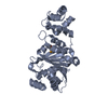
Protein
Nucleic acid
Ligand
Solvent
Total
Num. atoms
3876
0
0
236
4112
Refine LS restraints
Refine-ID
Type
Dev ideal
Dev ideal target
Number
X-RAY DIFFRACTION
r_bond_refined_d
0.015
0.022
3960
X-RAY DIFFRACTION
r_bond_other_d
X-RAY DIFFRACTION
r_angle_refined_deg
1.352
1.963
5395
X-RAY DIFFRACTION
r_angle_other_deg
X-RAY DIFFRACTION
r_dihedral_angle_1_deg
5.31
5
502
X-RAY DIFFRACTION
r_dihedral_angle_2_deg
X-RAY DIFFRACTION
r_chiral_restr
0.09
0.2
626
X-RAY DIFFRACTION
r_gen_planes_refined
0.005
0.02
3024
X-RAY DIFFRACTION
r_gen_planes_other
X-RAY DIFFRACTION
r_nbd_refined
0.208
0.2
1826
X-RAY DIFFRACTION
r_nbd_other
X-RAY DIFFRACTION
r_nbtor_other
X-RAY DIFFRACTION
r_xyhbond_nbd_refined
0.133
0.2
244
X-RAY DIFFRACTION
r_xyhbond_nbd_other
X-RAY DIFFRACTION
r_symmetry_vdw_refined
0.221
0.2
56
X-RAY DIFFRACTION
r_symmetry_vdw_other
X-RAY DIFFRACTION
r_symmetry_hbond_refined
0.126
0.2
16
X-RAY DIFFRACTION
r_symmetry_hbond_other
X-RAY DIFFRACTION
r_mcbond_it
0.714
1.5
2532
X-RAY DIFFRACTION
r_mcangle_it
1.315
2
4105
X-RAY DIFFRACTION
r_scbond_it
2.284
3
1428
X-RAY DIFFRACTION
r_scangle_it
3.666
4.5
1290
X-RAY DIFFRACTION
r_rigid_bond_restr
X-RAY DIFFRACTION
r_sphericity_free
X-RAY DIFFRACTION
r_sphericity_bonded
LS refinement shell
Resolution: 2.1→2.155 Å / Total num. of bins used: 20 /
Rfactor
Num. reflection
Rfree
0.279
209
Rwork
0.218
1829
Refinement TLS params.
Method: refined / Refine-ID: X-RAY DIFFRACTION
ID
L11 (°2)
L12 (°2)
L13 (°2)
L22 (°2)
L23 (°2)
L33 (°2)
S11 (Å °)
S12 (Å °)
S13 (Å °)
S21 (Å °)
S22 (Å °)
S23 (Å °)
S31 (Å °)
S32 (Å °)
S33 (Å °)
T11 (Å2)
T12 (Å2)
T13 (Å2)
T22 (Å2)
T23 (Å2)
T33 (Å2)
Origin x (Å)
Origin y (Å)
Origin z (Å)
1
0.9481
0.059
-0.0395
0.3725
-0.3619
1.7122
-0.0245
-0.0576
0.056
0.0469
-0.0706
-0.0458
-0.0805
-0.0012
0.095
0.0659
-0.0041
-0.0371
0.0642
-0.0035
0.0305
45.5337
25.5625
20.5766
2
2.9461
-0.105
-1.3134
0.72
-0.7213
2.5606
0.0104
-0.2106
0.0303
-0.0451
-0.148
-0.1833
-0.1259
0.3795
0.1376
0.0487
-0.0091
0
0.1158
0.0556
0.0471
62.9155
33.2071
-5.1564
3
1.6231
0.4465
-0.7527
0.5101
0.1813
0.8494
-0.0725
-0.0773
-0.1042
0.0305
-0.0108
-0.0364
-0.012
0.0403
0.0833
0.0564
0.0085
-0.0117
0.0013
-0.0018
0.0869
103.9478
41.8075
-39.1212
4
6.3308
1.1531
-3.3829
1.3317
-1.6333
4.7289
-0.4713
-0.561
-0.7072
0.1665
0.0644
-0.1994
0.2481
0.4179
0.4069
0.1011
0.0566
0.0893
0.0988
0.0674
0.1392
82.4455
29.4999
-19.4404
Refinement TLS group
ID
Refine-ID
Refine TLS-ID
Auth asym-ID
Label asym-ID
Auth seq-ID
Label seq-ID
1
X-RAY DIFFRACTION
1
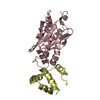
A
A
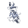
17 - 195
1 - 179
2
X-RAY DIFFRACTION
2
A
A
203 - 268
187 - 252
3
X-RAY DIFFRACTION
3
B
B
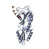
17 - 195
1 - 179
4
X-RAY DIFFRACTION
4
B
B
203 - 268
187 - 252
+
About Yorodumi
-
News
-
Feb 9, 2022. New format data for meta-information of EMDB entries
New format data for meta-information of EMDB entries
Version 3 of the EMDB header file is now the official format.
The previous official version 1.9 will be removed from the archive.
In the structure databanks used in Yorodumi, some data are registered as the other names, "COVID-19 virus" and "2019-nCoV". Here are the details of the virus and the list of structure data.
Jan 31, 2019. EMDB accession codes are about to change! (news from PDBe EMDB page)
EMDB accession codes are about to change! (news from PDBe EMDB page)
The allocation of 4 digits for EMDB accession codes will soon come to an end. Whilst these codes will remain in use, new EMDB accession codes will include an additional digit and will expand incrementally as the available range of codes is exhausted. The current 4-digit format prefixed with “EMD-” (i.e. EMD-XXXX) will advance to a 5-digit format (i.e. EMD-XXXXX), and so on. It is currently estimated that the 4-digit codes will be depleted around Spring 2019, at which point the 5-digit format will come into force.
The EM Navigator/Yorodumi systems omit the EMD- prefix.
Related info.:Q: What is EMD? / ID/Accession-code notation in Yorodumi/EM Navigator
Yorodumi is a browser for structure data from EMDB, PDB, SASBDB, etc.
This page is also the successor to EM Navigator detail page, and also detail information page/front-end page for Omokage search.
The word "yorodu" (or yorozu) is an old Japanese word meaning "ten thousand". "mi" (miru) is to see.
Related info.:EMDB / PDB / SASBDB / Comparison of 3 databanks / Yorodumi Search / Aug 31, 2016. New EM Navigator & Yorodumi / Yorodumi Papers / Jmol/JSmol / Function and homology information / Changes in new EM Navigator and Yorodumi
 Movie
Movie Controller

 Yorodumi
 Open data
 Basic information
 Components
 Keywords
 Function and homology information
 X-RAY DIFFRACTION /
  Authors
 Citation
 Structure visualization
 Downloads & links
 Other downloads
 PDBj
PDBj Assembly

 Mass: 18.015 Da / Num. of mol.: 236 / Source method: isolated from a natural source / Formula: H2O
 Sample preparation
 Processing