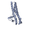
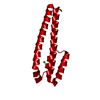
 Movie
Movie Controller


+ Open data

- Basic information
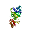
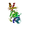
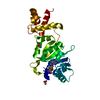
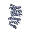
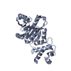
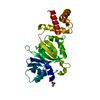
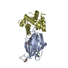
| Entry | Database: PDB / ID: 2erc | ||||||
|---|---|---|---|---|---|---|---|
| Title | CRYSTAL STRUCTURE OF ERMC' A RRNA-METHYL TRANSFERASE | ||||||
 Components Components | RRNA METHYL TRANSFERASE | ||||||
 Keywords Keywords | METHYLTRANSFERASE / ANTIBIOTIC RESISTANCE / RRNA METHYLTRANSFERASE / ERYTHROMYCIN RESISTANCE METHYLASE C' | ||||||
| Function / homology |  Function and homology information Function and homology information23S rRNA (adenine2085-N6)-dimethyltransferase / 23S rRNA (adenine(2085)-N(6))-dimethyltransferase activity / rRNA (adenine-N6,N6-)-dimethyltransferase activity / response to antibiotic / RNA binding / cytosol Similarity search - Function | ||||||
| Biological species |  | ||||||
| Method |  X-RAY DIFFRACTION / SYNCHROTRON / MAD PHASING IN A DIFFERENT EXPERIMENT / Resolution: 3.03 Å X-RAY DIFFRACTION / SYNCHROTRON / MAD PHASING IN A DIFFERENT EXPERIMENT / Resolution: 3.03 Å | ||||||
 Authors Authors | Bussiere, D.E. / Muchmore, S.W. / Dealwis, C.G. / Schluckebier, G. / Abad-Zapatero, C. | ||||||
 Citation Citation | Journal: Biochemistry / Year: 1998 Title: Crystal structure of ErmC', an rRNA methyltransferase which mediates antibiotic resistance in bacteria. Authors: Bussiere, D.E. / Muchmore, S.W. / Dealwis, C.G. / Schluckebier, G. / Nienaber, V.L. / Edalji, R.P. / Walter, K.A. / Ladror, U.S. / Holzman, T.F. / Abad-Zapatero, C. #1: Journal: J.Bacteriol. / Year: 1995Title: Substrate Requirements for Ermc' Methyltransferase Activity Authors: Zhong, P. / Pratt, S.D. / Edalji, R.P. / Walter, K.A. / Holzman, T.F. / Shivakumar, A.G. / Katz, L. #2: Journal: J.Bacteriol. / Year: 1986Title: Sequence and Properties of Pim13, a Macrolide-Lincosamide-Streptogramin B Resistance Plasmid from Bacillus Subtilis Authors: Monod, M. / Denoya, C. / Dubnau, D. | ||||||
| History |
|
- Structure visualization
| Structure viewer | Molecule: MolmilJmol/JSmol |
|---|
- Downloads & links
-Download
| PDBx/mmCIF format | 2erc.cif.gz | 107 KB | Display | PDBx/mmCIF format |
|---|---|---|---|---|
| PDB format | pdb2erc.ent.gz | 84.5 KB | Display | PDB format |
| PDBx/mmJSON format | 2erc.json.gz | Tree view | PDBx/mmJSON format | |
| Others |  Other downloads Other downloads |
-Validation report
| Arichive directory | https://data.pdbj.org/pub/pdb/validation_reports/er/2ercftp://data.pdbj.org/pub/pdb/validation_reports/er/2erc | HTTPS FTP |
|---|
-Related structure data
| Similar structure data |
|---|
-Links
 PDBj
PDBj- Assembly
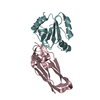
| Deposited unit | 
| ||||||||
|---|---|---|---|---|---|---|---|---|---|
| 1 | 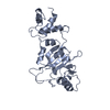
| ||||||||
| 2 | 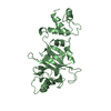
| ||||||||
| Unit cell |
| ||||||||
| Noncrystallographic symmetry (NCS) | NCS oper: (Code: given Matrix: (-0.4984, 0.8669, -0.0024), Vector: |
-Components
| #1: Protein | Mass: 28955.529 Da / Num. of mol.: 2 / Fragment: RESIDUES 10-244 Source method: isolated from a genetically manipulated source Source: (gene. exp.) |
|---|
-Experimental details
-Experiment
| Experiment | Method: X-RAY DIFFRACTION / Number of used crystals: 1 |
|---|
- Sample preparation
| Crystal | Density Matthews: 3.1 Å3/Da / Density % sol: 55 % Description: DATA FOR MAD PHASING WAS DONE WITH OTHER EXPERIMENTAL CONDITIONS. | |||||||||||||||||||||||||||||||||||||||||||||||||||||||||||||||
|---|---|---|---|---|---|---|---|---|---|---|---|---|---|---|---|---|---|---|---|---|---|---|---|---|---|---|---|---|---|---|---|---|---|---|---|---|---|---|---|---|---|---|---|---|---|---|---|---|---|---|---|---|---|---|---|---|---|---|---|---|---|---|---|---|
| Crystal grow | pH: 4 / Details: pH 4.0 | |||||||||||||||||||||||||||||||||||||||||||||||||||||||||||||||
| Crystal grow | *PLUS pH: 7.5 / Method: vapor diffusion, hanging drop | |||||||||||||||||||||||||||||||||||||||||||||||||||||||||||||||
| Components of the solutions | *PLUS
|
-Data collection
| Diffraction | Mean temperature: 103 K |
|---|---|
| Diffraction source | Source: SYNCHROTRON / Site: CHESS  / Beamline: F1 / Wavelength: 0.92178 / Beamline: F1 / Wavelength: 0.92178 |
| Detector | Type: TATE ET AL. (1995) J. APPL. CRYST. 28, 196-205. / Detector: CCD / Date: Oct 1, 1996 / Details: MIRROR |
| Radiation | Monochromator: SI(111) / Monochromatic (M) / Laue (L): M / Scattering type: x-ray |
| Radiation wavelength | Wavelength: 0.92178 Å / Relative weight: 1 |
| Reflection | Highest resolution: 3 Å / Num. obs: 14568 / % possible obs: 94 % / Observed criterion σ(I): 1 / Redundancy: 9.5 % / Rmerge(I) obs: 0.068 / Rsym value: 0.12 / Net I/σ(I): 10 |
| Reflection shell | Resolution: 3→3.5 Å / Redundancy: 6 % / Rmerge(I) obs: 0.027 / Mean I/σ(I) obs: 2 / Rsym value: 0.03 / % possible all: 84.3 |
| Reflection | *PLUS Num. measured all: 138382 |
| Reflection shell | *PLUS % possible obs: 84.3 % |
- Processing
| Software |
| ||||||||||||||||||||||||||||||||||||||||||||||||||||||||||||
|---|---|---|---|---|---|---|---|---|---|---|---|---|---|---|---|---|---|---|---|---|---|---|---|---|---|---|---|---|---|---|---|---|---|---|---|---|---|---|---|---|---|---|---|---|---|---|---|---|---|---|---|---|---|---|---|---|---|---|---|---|---|
| Refinement | Method to determine structure: MAD PHASING IN A DIFFERENT EXPERIMENT Resolution: 3.03→8 Å / Rfactor Rfree error: 0.01 / Data cutoff high absF: 10000000000000 / Data cutoff low absF: 0.0001 / Cross valid method: THROUGHOUT / σ(F): 0 Details: STRUCTURE WAS SOLVED IN P622 SPACE GROUP BUT WAS REFINED IN P6.
| ||||||||||||||||||||||||||||||||||||||||||||||||||||||||||||
| Displacement parameters | Biso mean: 67.4 Å2
| ||||||||||||||||||||||||||||||||||||||||||||||||||||||||||||
| Refine analyze |
| ||||||||||||||||||||||||||||||||||||||||||||||||||||||||||||
| Refinement step | Cycle: LAST / Resolution: 3.03→8 Å
| ||||||||||||||||||||||||||||||||||||||||||||||||||||||||||||
| Refine LS restraints |
| ||||||||||||||||||||||||||||||||||||||||||||||||||||||||||||
| Refine LS restraints NCS | NCS model details: RESTRAINTS / Rms dev Biso : 0.05 Å2 / Rms dev position: 0.04 Å / Weight Biso : 2 / Weight position: 200 | ||||||||||||||||||||||||||||||||||||||||||||||||||||||||||||
| LS refinement shell | Resolution: 3.03→3.13 Å / Rfactor Rfree error: 0.049 / Total num. of bins used: 10
| ||||||||||||||||||||||||||||||||||||||||||||||||||||||||||||
| Xplor file |
| ||||||||||||||||||||||||||||||||||||||||||||||||||||||||||||
| Software | *PLUS Name: X-PLOR / Version: 3.1 / Classification: refinement | ||||||||||||||||||||||||||||||||||||||||||||||||||||||||||||
| Refinement | *PLUS | ||||||||||||||||||||||||||||||||||||||||||||||||||||||||||||
| Solvent computation | *PLUS | ||||||||||||||||||||||||||||||||||||||||||||||||||||||||||||
| Displacement parameters | *PLUS | ||||||||||||||||||||||||||||||||||||||||||||||||||||||||||||
| Refine LS restraints | *PLUS
| ||||||||||||||||||||||||||||||||||||||||||||||||||||||||||||
| LS refinement shell | *PLUS Rfactor obs: 0.313 |
