 Movie
Movie Controller

[English] 日本語
 Yorodumi
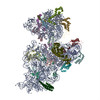
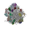
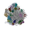
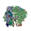
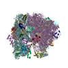
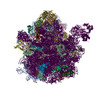
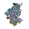
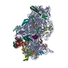
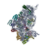
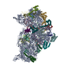
- PDB-4a2i: Cryo-electron Microscopy Structure of the 30S Subunit in Complex ... -
+ Open data

- Basic information
| Entry | Database: PDB / ID: 4a2i | ||||||
|---|---|---|---|---|---|---|---|
| Title | Cryo-electron Microscopy Structure of the 30S Subunit in Complex with the YjeQ Biogenesis Factor | ||||||
 Components Components |
| ||||||
 Keywords Keywords | RIBOSOME/HYDROLASE / RIBOSOME-HYDROLASE COMPLEX / RIBOSOME ASSEMBLY | ||||||
| Function / homology |  Function and homology information Function and homology informationtranscription antitermination factor activity, RNA binding / ornithine decarboxylase inhibitor activity / misfolded RNA binding / Group I intron splicing / RNA folding / Hydrolases; Acting on acid anhydrides; In phosphorus-containing anhydrides / four-way junction DNA binding / regulation of mRNA stability / negative regulation of translational initiation / mRNA regulatory element binding translation repressor activity ...transcription antitermination factor activity, RNA binding / ornithine decarboxylase inhibitor activity / misfolded RNA binding / Group I intron splicing / RNA folding / Hydrolases; Acting on acid anhydrides; In phosphorus-containing anhydrides / four-way junction DNA binding / regulation of mRNA stability / negative regulation of translational initiation / mRNA regulatory element binding translation repressor activity / positive regulation of RNA splicing / regulation of DNA-templated transcription elongation / transcription elongation factor complex / transcription antitermination / DNA endonuclease activity / DNA-templated transcription termination / maintenance of translational fidelity / mRNA 5'-UTR binding / regulation of translation / ribosomal small subunit assembly / ribosome biogenesis / ribosomal small subunit biogenesis / small ribosomal subunit / small ribosomal subunit rRNA binding / cytosolic small ribosomal subunit / cytoplasmic translation / tRNA binding / negative regulation of translation / rRNA binding / structural constituent of ribosome / ribosome / translation / response to antibiotic / hydrolase activity / mRNA binding / GTPase activity / GTP binding / RNA binding / zinc ion binding / membrane / metal ion binding / cytoplasm / cytosol Similarity search - Function | ||||||
| Biological species |  SALMONELLA ENTERICA SUBSP. ENTERICA SEROVAR TYPHIMURIUM (bacteria) SALMONELLA ENTERICA SUBSP. ENTERICA SEROVAR TYPHIMURIUM (bacteria) | ||||||
| Method | ELECTRON MICROSCOPY / single particle reconstruction / cryo EM / Resolution: 16.5 Å | ||||||
 Authors Authors | Jomaa, A. / Stewart, G. / Mears, J.A. / Kireeva, I. / Brown, E.D. / Ortega, J. | ||||||
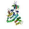
 Citation Citation | Journal: RNA / Year: 2011 Title: Cryo-electron microscopy structure of the 30S subunit in complex with the YjeQ biogenesis factor. Authors: Ahmad Jomaa / Geordie Stewart / Jason A Mears / Inga Kireeva / Eric D Brown / Joaquin Ortega /  Abstract: YjeQ is a protein broadly conserved in bacteria containing an N-terminal oligonucleotide/oligosaccharide fold (OB-fold) domain, a central GTPase domain, and a C-terminal zinc-finger domain. YjeQ ...YjeQ is a protein broadly conserved in bacteria containing an N-terminal oligonucleotide/oligosaccharide fold (OB-fold) domain, a central GTPase domain, and a C-terminal zinc-finger domain. YjeQ binds tightly and stoichiometrically to the 30S subunit, which stimulates its GTPase activity by 160-fold. Despite growing evidence for the involvement of the YjeQ protein in bacterial 30S subunit assembly, the specific function and mechanism of this protein remain unclear. Here, we report the costructure of YjeQ with the 30S subunit obtained by cryo-electron microscopy. The costructure revealed that YjeQ interacts simultaneously with helix 44, the head and the platform of the 30S subunit. This binding location of YjeQ in the 30S subunit suggests a chaperone role in processing of the 3' end of the rRNA as well as in mediating the correct orientation of the main domains of the 30S subunit. In addition, the YjeQ binding site partially overlaps with the interaction site of initiation factors 2 and 3, and upon binding, YjeQ covers three inter-subunit bridges that are important for the association of the 30S and 50S subunits. Hence, our structure suggests that YjeQ may assist in ribosome maturation by preventing premature formation of the translation initiation complex and association with the 50S subunit. Together, these results support a role for YjeQ in the late stages of 30S maturation. | ||||||
| History |
| ||||||
| Remark 700 | SHEET DETERMINATION METHOD: DSSP THE SHEETS PRESENTED AS "VA" IN EACH CHAIN ON SHEET RECORDS BELOW ... SHEET DETERMINATION METHOD: DSSP THE SHEETS PRESENTED AS "VA" IN EACH CHAIN ON SHEET RECORDS BELOW IS ACTUALLY AN 5-STRANDED BARREL THIS IS REPRESENTED BY A 6-STRANDED SHEET IN WHICH THE FIRST AND LAST STRANDS ARE IDENTICAL. |
- Structure visualization
| Movie |
Movie viewer |
|---|---|
| Structure viewer | Molecule: MolmilJmol/JSmol |
- Downloads & links
-Download
| PDBx/mmCIF format | 4a2i.cif.gz | 1.2 MB | Display | PDBx/mmCIF format |
|---|---|---|---|---|
| PDB format | pdb4a2i.ent.gz | 943.7 KB | Display | PDB format |
| PDBx/mmJSON format | 4a2i.json.gz | Tree view | PDBx/mmJSON format | |
| Others |  Other downloads Other downloads |
-Validation report
| Arichive directory | https://data.pdbj.org/pub/pdb/validation_reports/a2/4a2iftp://data.pdbj.org/pub/pdb/validation_reports/a2/4a2i | HTTPS FTP |
|---|
-Related structure data
| Related structure data | 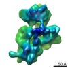 1895MC M: map data used to model this data C: citing same article ( |
|---|---|
| Similar structure data |


-Links
 PDBj
PDBj


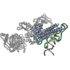
- Assembly
| Deposited unit | 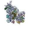
|
|---|---|
| 1 |
|
-Components
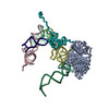
-30S RIBOSOMAL PROTEIN ... , 20 types, 20 molecules BCDEFGHIJKLMNOPQRSTU
| #2: Protein | Mass: 24253.943 Da / Num. of mol.: 1 / Source method: isolated from a natural source / Source: (natural) |
|---|---|
| #3: Protein | Mass: 23078.785 Da / Num. of mol.: 1 / Source method: isolated from a natural source / Source: (natural) |
| #4: Protein | Mass: 23383.002 Da / Num. of mol.: 1 / Source method: isolated from a natural source / Source: (natural) |
| #5: Protein | Mass: 15804.282 Da / Num. of mol.: 1 / Source method: isolated from a natural source / Source: (natural) |
| #6: Protein | Mass: 11669.371 Da / Num. of mol.: 1 / Source method: isolated from a natural source / Source: (natural) |
| #7: Protein | Mass: 16764.406 Da / Num. of mol.: 1 / Source method: isolated from a natural source / Source: (natural) |
| #8: Protein | Mass: 14015.361 Da / Num. of mol.: 1 / Source method: isolated from a natural source / Source: (natural) |
| #9: Protein | Mass: 14554.882 Da / Num. of mol.: 1 / Source method: isolated from a natural source / Source: (natural) |
| #10: Protein | Mass: 11196.988 Da / Num. of mol.: 1 / Source method: isolated from a natural source / Source: (natural) |
| #11: Protein | Mass: 12487.200 Da / Num. of mol.: 1 / Source method: isolated from a natural source / Source: (natural) |
| #12: Protein | Mass: 13636.961 Da / Num. of mol.: 1 / Source method: isolated from a natural source / Source: (natural) |
| #13: Protein | Mass: 12625.753 Da / Num. of mol.: 1 / Source method: isolated from a natural source / Source: (natural) |
| #14: Protein | Mass: 11475.364 Da / Num. of mol.: 1 / Source method: isolated from a natural source / Source: (natural) |
| #15: Protein | Mass: 10188.687 Da / Num. of mol.: 1 / Source method: isolated from a natural source / Source: (natural) |
| #16: Protein | Mass: 9207.572 Da / Num. of mol.: 1 / Source method: isolated from a natural source / Source: (natural) |
| #17: Protein | Mass: 9263.946 Da / Num. of mol.: 1 / Source method: isolated from a natural source / Source: (natural) |
| #18: Protein | Mass: 6466.477 Da / Num. of mol.: 1 / Source method: isolated from a natural source / Source: (natural) |
| #19: Protein | Mass: 9057.626 Da / Num. of mol.: 1 / Source method: isolated from a natural source / Source: (natural) |
| #20: Protein | Mass: 9506.190 Da / Num. of mol.: 1 / Source method: isolated from a natural source / Source: (natural) |
| #21: Protein | Mass: 6067.081 Da / Num. of mol.: 1 / Source method: isolated from a natural source / Source: (natural) |
-RNA chain / Protein , 2 types, 2 molecules AV
| #1: RNA chain | Mass: 495927.719 Da / Num. of mol.: 1 / Source method: isolated from a natural source / Source: (natural) |
|---|---|
| #22: Protein | Mass: 30816.074 Da / Num. of mol.: 1 Source method: isolated from a genetically manipulated source Source: (gene. exp.) SALMONELLA ENTERICA SUBSP. ENTERICA SEROVAR TYPHIMURIUM (bacteria)Plasmid: PDEST17 / Production host: |
-Experimental details
-Experiment
| Experiment | Method: ELECTRON MICROSCOPY |
|---|---|
| EM experiment | Aggregation state: PARTICLE / 3D reconstruction method: single particle reconstruction |
- Sample preparation
| Component | Name: ESCHERICHIA COLI 30S RIBOSOMAL SUBUNIT WITH YJEQ PROTEIN BOUND IN THE PRESENCE OF GMP-PNP Type: RIBOSOME |
|---|---|
| Buffer solution | Name: 10 MM TRIS-HCL PH 7.5, 10 MM MAGNESIUM ACETATE, 60 MM NH4CL, 3 MM 2- MERCAPTOETHANOL AND 2 MM GMP-PNP pH: 7.5 Details: 10 MM TRIS-HCL PH 7.5, 10 MM MAGNESIUM ACETATE, 60 MM NH4CL, 3 MM 2- MERCAPTOETHANOL AND 2 MM GMP-PNP |
| Specimen | Conc.: 0.9 mg/ml / Embedding applied: NO / Shadowing applied: NO / Staining applied: NO / Vitrification applied: YES |
| Specimen support | Details: FORMVAR PLUS CARBON |
| Vitrification | Instrument: FEI VITROBOT MARK III / Cryogen name: ETHANE / Details: BLOT FOR 7 SECONDS IN FEI VITROBOT III |
- Electron microscopy imaging
| Microscopy | Model: JEOL 2010F / Date: May 12, 2010 |
|---|---|
| Electron gun | Electron source:  FIELD EMISSION GUN / Accelerating voltage: 200 kV / Illumination mode: FLOOD BEAM FIELD EMISSION GUN / Accelerating voltage: 200 kV / Illumination mode: FLOOD BEAM |
| Electron lens | Mode: BRIGHT FIELD / Nominal magnification: 50000 X / Calibrated magnification: 50000 X / Nominal defocus max: 3900 nm / Nominal defocus min: 650 nm / Cs: 1 mm |
| Specimen holder | Temperature: 100 K / Tilt angle min: 0 ° |
| Image recording | Electron dose: 15 e/Å2 / Film or detector model: KODAK SO-163 FILM |
| Image scans | Num. digital images: 150 |
| Radiation wavelength | Relative weight: 1 |
- Processing
| EM software |
| |||||||||||||||||||||
|---|---|---|---|---|---|---|---|---|---|---|---|---|---|---|---|---|---|---|---|---|---|---|
| CTF correction | Details: EACH MICROGRAPH | |||||||||||||||||||||
| Symmetry | Point symmetry: C1 (asymmetric) | |||||||||||||||||||||
| 3D reconstruction | Method: PROJECTION MATCHING / Resolution: 16.5 Å / Num. of particles: 16228 / Nominal pixel size: 2.54 Å / Actual pixel size: 2.54 Å Details: THE COORDINATES IN THIS ENTRY WERE GENERATED BY MANUAL DOCKING OF THE STRUCTURE OF THE ESCHERICHIA COLI 30S RIBOSOMAL SUBUNIT (2AVY) AND SALMONELLA TYPHYMURIUM (2RCN) INTO THE DENSITY MAP OF ...Details: THE COORDINATES IN THIS ENTRY WERE GENERATED BY MANUAL DOCKING OF THE STRUCTURE OF THE ESCHERICHIA COLI 30S RIBOSOMAL SUBUNIT (2AVY) AND SALMONELLA TYPHYMURIUM (2RCN) INTO THE DENSITY MAP OF THE ESCHERICHIA COLI 30S_YJEQ COMPLEX GENERATED BY CRYO-ELECTRON MICROSCOPY. THE YEJQ PROTEIN WAS FITTED AS THREE SEPARATE DOMAINS: THE OB-FOLD, THE GTPASE DOMAIN, AND THE ZINC-FINGER DOMAIN. THE PROTEIN DATA BANK CONVENTIONS REQUIRE TO ENTER INFORMATION ABOUT THE UNIT CELL, CRYSTAL DATA AND COORDINATE SYSTEM. THESE INFORMATION IS MEANINGLESS IN THIS ENTRY. SUBMISSION BASED ON EXPERIMENTAL DATA FROM EMDB EMD-1895. Symmetry type: POINT | |||||||||||||||||||||
| Atomic model building | Protocol: OTHER / Space: REAL / Target criteria: Cross-correlation coefficient / Details: METHOD--MANUAL REFINEMENT PROTOCOL--X-RAY | |||||||||||||||||||||
| Atomic model building |
| |||||||||||||||||||||
| Refinement | Highest resolution: 16.5 Å | |||||||||||||||||||||
| Refinement step | Cycle: LAST / Highest resolution: 16.5 Å
|

