[English] 日本語
 Yorodumi
- PDB-1nkw: Crystal Structure Of The Large Ribosomal Subunit From Deinococcus... -
+ Open data
- Basic information
| Entry | Database: PDB / ID: 1nkw | |||||||||
|---|---|---|---|---|---|---|---|---|---|---|
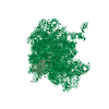
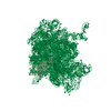
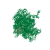
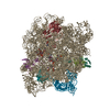
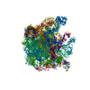
| Title | Crystal Structure Of The Large Ribosomal Subunit From Deinococcus Radiodurans | |||||||||
 Components Components |
| |||||||||
 Keywords Keywords | RIBOSOME / large subunit / 50S / Deinococcus Radiodurans / X-ray structure / peptidyl-transferase / peptide bond formation | |||||||||
| Function / homology |  Function and homology information Function and homology informationlarge ribosomal subunit / transferase activity / 5S rRNA binding / ribosomal large subunit assembly / large ribosomal subunit rRNA binding / cytosolic large ribosomal subunit / cytoplasmic translation / tRNA binding / negative regulation of translation / rRNA binding ...large ribosomal subunit / transferase activity / 5S rRNA binding / ribosomal large subunit assembly / large ribosomal subunit rRNA binding / cytosolic large ribosomal subunit / cytoplasmic translation / tRNA binding / negative regulation of translation / rRNA binding / structural constituent of ribosome / ribosome / translation / ribonucleoprotein complex / mRNA binding / RNA binding / zinc ion binding / metal ion binding / cytoplasm Similarity search - Function | |||||||||
| Biological species |  Deinococcus radiodurans (radioresistant) Deinococcus radiodurans (radioresistant) | |||||||||
| Method |  X-RAY DIFFRACTION / X-RAY DIFFRACTION /  SYNCHROTRON / COMBINATION OF molecular replacement, SYNCHROTRON / COMBINATION OF molecular replacement,  MIR / Resolution: 3.1 Å MIR / Resolution: 3.1 Å | |||||||||
 Authors Authors | Harms, J.M. / Schluenzen, F. / Zarivach, R. / Bashan, A. / Gat, S. / Agmon, I. / Bartels, H. / Franceschi, F. / Yonath, A. | |||||||||
 Citation Citation |  Journal: Cell / Year: 2001 Journal: Cell / Year: 2001Title: High resolution structure of the large ribosomal subunit from a mesophilic eubacterium. Authors: J Harms / F Schluenzen / R Zarivach / A Bashan / S Gat / I Agmon / H Bartels / F Franceschi / A Yonath /  Abstract: We describe the high resolution structure of the large ribosomal subunit from Deinococcus radiodurans (D50S), a gram-positive mesophile suitable for binding of antibiotics and functionally relevant ...We describe the high resolution structure of the large ribosomal subunit from Deinococcus radiodurans (D50S), a gram-positive mesophile suitable for binding of antibiotics and functionally relevant ligands. The over-all structure of D50S is similar to that from the archae bacterium Haloarcula marismortui (H50S); however, a detailed comparison revealed significant differences, for example, in the orientation of nucleotides in peptidyl transferase center and in the structures of many ribosomal proteins. Analysis of ribosomal features involved in dynamic aspects of protein biosynthesis that are partially or fully disordered in H50S revealed the conformations of intersubunit bridges in unbound subunits, suggesting how they may change upon subunit association and how movements of the L1-stalk may facilitate the exit of tRNA. #1:  Journal: Nature / Year: 2001 Journal: Nature / Year: 2001Title: Structural basis for the interaction of antibiotics with the peptidyl transferase centre in eubacteria Authors: Schluenzen, F. / Zarivach, R. / Harms, J.M. / Bashan, A. / Tocilj, A. / Albrecht, R. / Yonath, A. / Franceschi, F. | |||||||||
| History |
|
- Structure visualization
| Structure viewer | Molecule:  Molmil Molmil Jmol/JSmol Jmol/JSmol |
|---|
- Downloads & links
- Download
| PDBx/mmCIF format |  1nkw.cif.gz 1nkw.cif.gz | 1.4 MB | Display |  PDBx/mmCIF format PDBx/mmCIF format |
|---|---|---|---|---|
| PDB format |  pdb1nkw.ent.gz pdb1nkw.ent.gz | 1.1 MB | Display |  PDB format PDB format |
| PDBx/mmJSON format |  1nkw.json.gz 1nkw.json.gz | Tree view |  PDBx/mmJSON format PDBx/mmJSON format | |
| Others |  Other downloads Other downloads |
-Validation report
| Arichive directory |  https://data.pdbj.org/pub/pdb/validation_reports/nk/1nkw https://data.pdbj.org/pub/pdb/validation_reports/nk/1nkw ftp://data.pdbj.org/pub/pdb/validation_reports/nk/1nkw ftp://data.pdbj.org/pub/pdb/validation_reports/nk/1nkw | HTTPS FTP |
|---|
-Related structure data
| Related structure data | 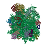 1ffkS S: Starting model for refinement |
|---|---|
| Similar structure data |
- Links
Links
- Assembly
| Deposited unit | 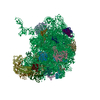
| ||||||||||
|---|---|---|---|---|---|---|---|---|---|---|---|
| 1 |
| ||||||||||
| Unit cell |
|
- Components
-RNA chain , 2 types, 2 molecules 09
| #1: RNA chain | Mass: 933405.000 Da / Num. of mol.: 1 / Source method: isolated from a natural source / Source: (natural)  Deinococcus radiodurans (radioresistant) / References: GenBank: 6460405 Deinococcus radiodurans (radioresistant) / References: GenBank: 6460405 |
|---|---|
| #2: RNA chain | Mass: 39911.859 Da / Num. of mol.: 1 / Source method: isolated from a natural source / Source: (natural)  Deinococcus radiodurans (radioresistant) / References: GenBank: 6460405 Deinococcus radiodurans (radioresistant) / References: GenBank: 6460405 |
+50S ribosomal protein ... , 28 types, 28 molecules ABCDEFGHIJKLMNOPQRSUWXYZ1234
-Protein , 1 types, 1 molecules T
| #22: Protein | Mass: 27004.121 Da / Num. of mol.: 1 / Source method: isolated from a natural source / Source: (natural)  Deinococcus radiodurans (radioresistant) / References: UniProt: Q9RX88 Deinococcus radiodurans (radioresistant) / References: UniProt: Q9RX88 |
|---|
-Experimental details
-Experiment
| Experiment | Method:  X-RAY DIFFRACTION / Number of used crystals: 1 X-RAY DIFFRACTION / Number of used crystals: 1 |
|---|
- Sample preparation
| Crystal | Density Matthews: 4 Å3/Da / Density % sol: 64 % | ||||||||||||||||||||||||||||||||||||||||||||||||||||||||||||||||||||||||||||||||||||
|---|---|---|---|---|---|---|---|---|---|---|---|---|---|---|---|---|---|---|---|---|---|---|---|---|---|---|---|---|---|---|---|---|---|---|---|---|---|---|---|---|---|---|---|---|---|---|---|---|---|---|---|---|---|---|---|---|---|---|---|---|---|---|---|---|---|---|---|---|---|---|---|---|---|---|---|---|---|---|---|---|---|---|---|---|---|
| Crystal grow | Method: vapor diffusion, sitting drop Details: ETHANOL, DIMETHYLHEXANEDIOL, MGCL2, KCL, HEPES, NH4CL, VAPOR DIFFUSION, SITTING DROP | ||||||||||||||||||||||||||||||||||||||||||||||||||||||||||||||||||||||||||||||||||||
| Components of the solutions |
| ||||||||||||||||||||||||||||||||||||||||||||||||||||||||||||||||||||||||||||||||||||
| Crystal grow | *PLUS Temperature: 18 ℃ / pH: 7.8 / Method: vapor diffusion | ||||||||||||||||||||||||||||||||||||||||||||||||||||||||||||||||||||||||||||||||||||
| Components of the solutions | *PLUS
|
-Data collection
| Diffraction | Mean temperature: 90 K |
|---|---|
| Diffraction source | Source:  SYNCHROTRON / Site: SYNCHROTRON / Site:  ESRF ESRF  / Beamline: ID14-4 / Wavelength: 0.9393 / Beamline: ID14-4 / Wavelength: 0.9393 |
| Detector | Type: ADSC QUANTUM 4 / Detector: CCD / Date: Apr 1, 2001 |
| Radiation | Monochromator: SI111 OR SI311 / Protocol: SINGLE WAVELENGTH / Monochromatic (M) / Laue (L): M / Scattering type: x-ray |
| Radiation wavelength | Wavelength: 0.9393 Å / Relative weight: 1 |
| Reflection | Resolution: 3→50 Å / Num. all: 441570 / Num. obs: 441570 / % possible obs: 95 % / Observed criterion σ(I): 0 |
| Reflection shell | Resolution: 3→3.05 Å / % possible all: 48.5 |
| Reflection | *PLUS Highest resolution: 3 Å / Lowest resolution: 50 Å / Num. measured all: 4787954 / Rmerge(I) obs: 0.145 |
| Reflection shell | *PLUS Highest resolution: 3 Å / % possible obs: 48.5 % / Rmerge(I) obs: 0.44 / Mean I/σ(I) obs: 1.8 |
- Processing
| Software |
| |||||||||||||||||||||||||||
|---|---|---|---|---|---|---|---|---|---|---|---|---|---|---|---|---|---|---|---|---|---|---|---|---|---|---|---|---|
| Refinement | Method to determine structure: COMBINATION OF molecular replacement,  MIR MIRStarting model: PDB ENTRY 1FFK Resolution: 3.1→15 Å / Rfactor Rfree error: 0.003 / Cross valid method: THROUGHOUT / σ(F): 0 / Stereochemistry target values: ENGH & HUBER
| |||||||||||||||||||||||||||
| Refinement step | Cycle: LAST / Resolution: 3.1→15 Å
| |||||||||||||||||||||||||||
| Refine LS restraints |
| |||||||||||||||||||||||||||
| Refinement | *PLUS | |||||||||||||||||||||||||||
| Solvent computation | *PLUS | |||||||||||||||||||||||||||
| Displacement parameters | *PLUS |
 Movie
Movie Controller





 PDBj
PDBj


