Monochromator: Si 111 / Protocol: SINGLE WAVELENGTH / Monochromatic (M) / Laue (L): M / Scattering type: x-ray
Radiation wavelength
Wavelength: 0.97929 Å / Relative weight: 1
Reflection
Resolution: 3.66→30 Å / Num. obs: 243598 / % possible obs: 93.1 % / Redundancy: 8.1 % / Rmerge(I) obs: 0.19 / Net I/σ(I): 9.1
Reflection shell
Resolution: 3.66→3.79 Å / Redundancy: 3.7 % / Rmerge(I) obs: 0.791 / Mean I/σ(I) obs: 1.4 / % possible all: 70
-
Processing
Software
Name
Version
Classification
CNS
1.1
refinement
HKL-2000
datareduction
SCALEPACK
datascaling
Refinement
Method to determine structure: FOURIER SYNTHESIS / Resolution: 3.66→29.96 Å / Rfactor Rfree error: 0.004 / Data cutoff high absF: 12688274.59 / Data cutoff low absF: 0 / Isotropic thermal model: RESTRAINED / Cross valid method: THROUGHOUT / σ(F): 0 / Details: the protein of this entry contains CA only
Rfactor
Num. reflection
% reflection
Selection details
Rfree
0.334
12167
5 %
RANDOM
Rwork
0.26
-
-
-
obs
-
243559
93 %
-
Displacement parameters
Baniso -1
Baniso -2
Baniso -3
1-
14.126 Å2
0 Å2
0 Å2
2-
-
-49.692 Å2
0 Å2
3-
-
-
35.566 Å2
Refinement step
Cycle: LAST / Resolution: 3.66→29.96 Å
Protein
Nucleic acid
Ligand
Solvent
Total
Num. atoms
205
59336
36
0
59577
Refine LS restraints
Refine-ID
Type
Dev ideal
X-RAY DIFFRACTION
c_bond_d
0.008
X-RAY DIFFRACTION
c_bond_d_na
X-RAY DIFFRACTION
c_bond_d_prot
X-RAY DIFFRACTION
c_angle_d
X-RAY DIFFRACTION
c_angle_d_na
X-RAY DIFFRACTION
c_angle_d_prot
X-RAY DIFFRACTION
c_angle_deg
1.4
X-RAY DIFFRACTION
c_angle_deg_na
X-RAY DIFFRACTION
c_angle_deg_prot
X-RAY DIFFRACTION
c_dihedral_angle_d
X-RAY DIFFRACTION
c_dihedral_angle_d_na
X-RAY DIFFRACTION
c_dihedral_angle_d_prot
X-RAY DIFFRACTION
c_improper_angle_d
X-RAY DIFFRACTION
c_improper_angle_d_na
X-RAY DIFFRACTION
c_improper_angle_d_prot
X-RAY DIFFRACTION
c_mcbond_it
X-RAY DIFFRACTION
c_mcangle_it
X-RAY DIFFRACTION
c_scbond_it
X-RAY DIFFRACTION
c_scangle_it
+
About Yorodumi
-
News
-
Feb 9, 2022. New format data for meta-information of EMDB entries
New format data for meta-information of EMDB entries
Version 3 of the EMDB header file is now the official format.
The previous official version 1.9 will be removed from the archive.
In the structure databanks used in Yorodumi, some data are registered as the other names, "COVID-19 virus" and "2019-nCoV". Here are the details of the virus and the list of structure data.
Jan 31, 2019. EMDB accession codes are about to change! (news from PDBe EMDB page)
EMDB accession codes are about to change! (news from PDBe EMDB page)
The allocation of 4 digits for EMDB accession codes will soon come to an end. Whilst these codes will remain in use, new EMDB accession codes will include an additional digit and will expand incrementally as the available range of codes is exhausted. The current 4-digit format prefixed with “EMD-” (i.e. EMD-XXXX) will advance to a 5-digit format (i.e. EMD-XXXXX), and so on. It is currently estimated that the 4-digit codes will be depleted around Spring 2019, at which point the 5-digit format will come into force.
The EM Navigator/Yorodumi systems omit the EMD- prefix.
Related info.:Q: What is EMD? / ID/Accession-code notation in Yorodumi/EM Navigator
Yorodumi is a browser for structure data from EMDB, PDB, SASBDB, etc.
This page is also the successor to EM Navigator detail page, and also detail information page/front-end page for Omokage search.
The word "yorodu" (or yorozu) is an old Japanese word meaning "ten thousand". "mi" (miru) is to see.
Related info.:EMDB / PDB / SASBDB / Comparison of 3 databanks / Yorodumi Search / Aug 31, 2016. New EM Navigator & Yorodumi / Yorodumi Papers / Jmol/JSmol / Function and homology information / Changes in new EM Navigator and Yorodumi
 Movie
Movie Controller

 Yorodumi
 Open data
 Basic information
 Components
 Keywords
 Function and homology information
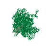
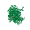
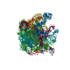
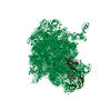
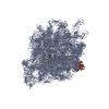
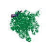
 Deinococcus radiodurans (radioresistant)
 X-RAY DIFFRACTION /
  Authors
 Citation
 Structure visualization
 Downloads & links
 Other downloads


 PDBj
PDBj


 Assembly
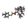
 Mass: 517.763 Da / Num. of mol.: 1 / Source method: obtained synthetically / Formula: C30H47NO4S
 Sample preparation
 / Beamline: 19-ID / Wavelength: 0.97929 Å
 Processing