 Movie
Movie Controller

[English] 日本語
 Yorodumi
- PDB-1njo: The crystal structure of the 50S Large ribosomal subunit from Dei... -
+ Open data

- Basic information
| Entry | Database: PDB / ID: 1njo | ||||||
|---|---|---|---|---|---|---|---|
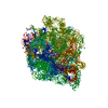
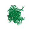
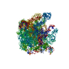
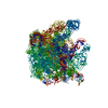
| Title | The crystal structure of the 50S Large ribosomal subunit from Deinococcus radiodurans complexed with a short substrate analog ACCPuromycin (ACCP) | ||||||
 Components Components |
| ||||||
 Keywords Keywords | RIBOSOME / Ribosomes / tRNA / puromycin / sparsomycin / peptidyl-transferase / peptide bond formation | ||||||
| Function / homology | : / RNA / RNA (> 10) / RNA (> 100) / RNA (> 1000) Function and homology information Function and homology information | ||||||
| Biological species |  Deinococcus radiodurans (radioresistant) Deinococcus radiodurans (radioresistant) | ||||||
| Method |  X-RAY DIFFRACTION / SYNCHROTRON / DIFFERENCE FOURIER MAPS / Resolution: 3.7 Å X-RAY DIFFRACTION / SYNCHROTRON / DIFFERENCE FOURIER MAPS / Resolution: 3.7 Å | ||||||
 Authors Authors | Bashan, A. / Agmon, I. / Zarivatch, R. / Schluenzen, F. / Harms, J.M. / Berisio, R. / Bartels, H. / Hansen, H.A. / Yonath, A. | ||||||
 Citation Citation | Journal: Mol.Cell / Year: 2003 Title: Structural basis of the ribosomal machinery for Peptide bond formation, translocation, and nascent chain progression Authors: Bashan, A. / Agmon, I. / Zarivatch, R. / Schluenzen, F. / Harms, J.M. / Berisio, R. / Bartels, H. / Franceschi, F. / Auerbach, T. / Hansen, H.A. / Kossoy, E. / Kessler, M. / Yonath, A. | ||||||
| History |
|
- Structure visualization
| Structure viewer | Molecule: MolmilJmol/JSmol |
|---|
- Downloads & links
-Download
| PDBx/mmCIF format | 1njo.cif.gz | 1.3 MB | Display | PDBx/mmCIF format |
|---|---|---|---|---|
| PDB format | pdb1njo.ent.gz | 1003.5 KB | Display | PDB format |
| PDBx/mmJSON format | 1njo.json.gz | Tree view | PDBx/mmJSON format | |
| Others |  Other downloads Other downloads |
-Validation report
| Arichive directory | https://data.pdbj.org/pub/pdb/validation_reports/nj/1njoftp://data.pdbj.org/pub/pdb/validation_reports/nj/1njo | HTTPS FTP |
|---|
-Related structure data
| Related structure data |  1njmC  1njnC  1njpC  1nkwS C: citing same article ( S: Starting model for refinement |
|---|---|
| Similar structure data |


-Links
 PDBj
PDBj


- Assembly
| Deposited unit | 
| ||||||||||
|---|---|---|---|---|---|---|---|---|---|---|---|
| 1 |
| ||||||||||
| Unit cell |
|
-Components
| #1: RNA chain | Mass: 933405.000 Da / Num. of mol.: 1 / Source method: isolated from a natural source / Source: (natural) Deinococcus radiodurans (radioresistant) / References: GenBank: 6460405 |
|---|---|
| #2: RNA chain | Mass: 1428.086 Da / Num. of mol.: 1 / Source method: obtained synthetically Details: RNA chain of the sequence ACC. The terminal C was coupled via a phosphodiester bond to the 5 OH of the N6-dimethyl moiety of puromycin |
-Experimental details
-Experiment
| Experiment | Method: X-RAY DIFFRACTION / Number of used crystals: 1 |
|---|
- Sample preparation
| Crystal | Density Matthews: 4 Å3/Da / Density % sol: 64 % | ||||||||||||||||||||||||||||||||||||||||||||||||||||||||||||||||||||||||||||||||||||
|---|---|---|---|---|---|---|---|---|---|---|---|---|---|---|---|---|---|---|---|---|---|---|---|---|---|---|---|---|---|---|---|---|---|---|---|---|---|---|---|---|---|---|---|---|---|---|---|---|---|---|---|---|---|---|---|---|---|---|---|---|---|---|---|---|---|---|---|---|---|---|---|---|---|---|---|---|---|---|---|---|---|---|---|---|---|
| Crystal grow | pH: 7.8 Details: ETHANOL, DIMETHYLHEXANEDIOL, MGCL2, KCL, HEPES, NH4CL; SOAKING CRYSTALS IN: 0.0125mM ACCP, pH 7.80 | ||||||||||||||||||||||||||||||||||||||||||||||||||||||||||||||||||||||||||||||||||||
| Components of the solutions |
| ||||||||||||||||||||||||||||||||||||||||||||||||||||||||||||||||||||||||||||||||||||
| Crystal grow | *PLUS Temperature: 18 ℃ / pH: 7.8 / Method: vapor diffusionDetails: Harms, J.M., (2001) Cell (Cambridge,Mass.), 107, 679. | ||||||||||||||||||||||||||||||||||||||||||||||||||||||||||||||||||||||||||||||||||||
| Components of the solutions | *PLUS
|
-Data collection
| Diffraction | Mean temperature: 90 K |
|---|---|
| Diffraction source | Source: SYNCHROTRON / Site: ESRF  / Beamline: ID14-4 / Wavelength: 0.9762 / Beamline: ID14-4 / Wavelength: 0.9762 |
| Detector | Type: ADSC QUANTUM 4 / Detector: CCD / Date: Feb 1, 2001 |
| Radiation | Protocol: SINGLE WAVELENGTH / Monochromatic (M) / Laue (L): M / Scattering type: x-ray |
| Radiation wavelength | Wavelength: 0.9762 Å / Relative weight: 1 |
| Reflection | Resolution: 3.5→50 Å / Num. obs: 249369 / % possible obs: 97.8 % / Observed criterion σ(I): -3 / Rsym value: 0.156 / Net I/σ(I): 7.9 |
| Reflection shell | Resolution: 3.7→3.76 Å / Mean I/σ(I) obs: 1.8 / Rsym value: 0.393 / % possible all: 97.3 |
| Reflection | *PLUS Highest resolution: 3.7 Å / Lowest resolution: 50 Å / Rmerge(I) obs: 0.156 |
| Reflection shell | *PLUS % possible obs: 97.3 % / Rmerge(I) obs: 0.393 |
- Processing
| Software |
| ||||||||||||||||||||||||||||||
|---|---|---|---|---|---|---|---|---|---|---|---|---|---|---|---|---|---|---|---|---|---|---|---|---|---|---|---|---|---|---|---|
| Refinement | Method to determine structure: DIFFERENCE FOURIER MAPS Starting model: PDB ENTRY 1NKW Resolution: 3.7→15 Å / Rfactor Rfree error: 0.003 / Cross valid method: THROUGHOUT / σ(F): 0 / Stereochemistry target values: ENGH & HUBER
| ||||||||||||||||||||||||||||||
| Refinement step | Cycle: LAST / Resolution: 3.7→15 Å
| ||||||||||||||||||||||||||||||
| Refine LS restraints |
| ||||||||||||||||||||||||||||||
| Refinement | *PLUS Rfactor Rfree: 0.305 | ||||||||||||||||||||||||||||||
| Solvent computation | *PLUS | ||||||||||||||||||||||||||||||
| Displacement parameters | *PLUS | ||||||||||||||||||||||||||||||
| Refine LS restraints | *PLUS Type: c_angle_d |
