 Movie
Movie Controller

[English] 日本語
 Yorodumi
- PDB-4n6s: Crystals of cross-linked stabilized and functional Phycobilisomes... -
+ Open data

- Basic information
| Entry | Database: PDB / ID: 4n6s | ||||||
|---|---|---|---|---|---|---|---|
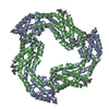
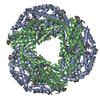
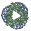
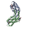
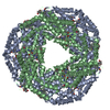
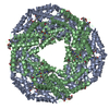
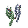
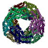
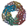
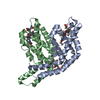
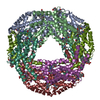
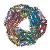
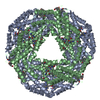
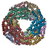
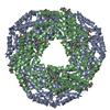
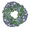
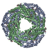
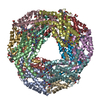
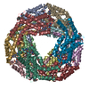
| Title | Crystals of cross-linked stabilized and functional Phycobilisomes: only phycocyanin rods contribute to diffraction. | ||||||
 Components Components |
| ||||||
 Keywords Keywords | PHOTOSYNTHESIS / antenna / glutaraldehyde cross-links / membrane associated | ||||||
| Function / homology |  Function and homology information Function and homology informationphycobilisome / plasma membrane-derived thylakoid membrane / photosynthesis Similarity search - Function | ||||||
| Biological species |  Thermosynechococcus vulcanus (bacteria) Thermosynechococcus vulcanus (bacteria) | ||||||
| Method |  X-RAY DIFFRACTION / SYNCHROTRON / MOLECULAR REPLACEMENT / Resolution: 2.4 Å X-RAY DIFFRACTION / SYNCHROTRON / MOLECULAR REPLACEMENT / Resolution: 2.4 Å | ||||||
 Authors Authors | David, L. / Prado, M. / Arteni, A. / Elmlund, D.A. / Blankenship, R.E. / Adir, N. | ||||||
 Citation Citation | Journal: Biochim.Biophys.Acta / Year: 2014 Title: Structural studies show energy transfer within stabilized phycobilisomes independent of the mode of rod-core assembly. Authors: David, L. / Prado, M. / Arteni, A.A. / Elmlund, D.A. / Blankenship, R.E. / Adir, N. | ||||||
| History |
|
- Structure visualization
| Structure viewer | Molecule: MolmilJmol/JSmol |
|---|
- Downloads & links
-Download
| PDBx/mmCIF format | 4n6s.cif.gz | 81.1 KB | Display | PDBx/mmCIF format |
|---|---|---|---|---|
| PDB format | pdb4n6s.ent.gz | 60.8 KB | Display | PDB format |
| PDBx/mmJSON format | 4n6s.json.gz | Tree view | PDBx/mmJSON format | |
| Others |  Other downloads Other downloads |
-Validation report
| Arichive directory | https://data.pdbj.org/pub/pdb/validation_reports/n6/4n6sftp://data.pdbj.org/pub/pdb/validation_reports/n6/4n6s | HTTPS FTP |
|---|
-Related structure data
| Related structure data | 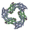 3o18S S: Starting model for refinement |
|---|---|
| Similar structure data |


-Links
 PDBj
PDBj- Assembly
| Deposited unit | 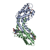
| ||||||||
|---|---|---|---|---|---|---|---|---|---|
| 1 | x 6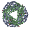
| ||||||||
| 2 | 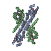
| ||||||||
| 3 |
| ||||||||
| Unit cell |
| ||||||||
| Details | THE AUTHOR STATES THAT THE BIOLOGICAL UNIT OF THIS PROTEIN IS UNKNOWN. |
-Components
| #1: Protein | Mass: 17470.656 Da / Num. of mol.: 1 / Source method: isolated from a natural source / Source: (natural) Thermosynechococcus vulcanus (bacteria) / References: UniProt: Q9AM02 | ||
|---|---|---|---|
| #2: Protein | Mass: 18216.652 Da / Num. of mol.: 1 / Source method: isolated from a natural source / Source: (natural) Thermosynechococcus vulcanus (bacteria) / References: UniProt: Q71RW8 | ||
| #3: Chemical |   Mass: 588.694 Da / Num. of mol.: 3 / Source method: obtained synthetically / Formula: C33H40N4O6 Mass: 588.694 Da / Num. of mol.: 3 / Source method: obtained synthetically / Formula: C33H40N4O6#4: Water | ChemComp-HOH / |  Mass: 18.015 Da / Num. of mol.: 110 / Source method: isolated from a natural source / Formula: H2O Mass: 18.015 Da / Num. of mol.: 110 / Source method: isolated from a natural source / Formula: H2O |
-Experimental details
-Experiment
| Experiment | Method: X-RAY DIFFRACTION / Number of used crystals: 1 |
|---|
- Sample preparation
| Crystal | Density Matthews: 2.88 Å3/Da / Density % sol: 57.28 % |
|---|---|
| Crystal grow | Temperature: 293 K / Method: vapor diffusion, hanging drop / pH: 7 Details: 10-15 mg/ml phycobilisome in 0.9M phosphate buffer against a resevoir of 1.2-1.4M phosphate buffer. , pH 7.0, VAPOR DIFFUSION, HANGING DROP, temperature 293K |
-Data collection
| Diffraction | Mean temperature: 100 K |
|---|---|
| Diffraction source | Source: SYNCHROTRON / Site: ESRF  / Beamline: ID23-1 / Wavelength: 0.9795 Å / Beamline: ID23-1 / Wavelength: 0.9795 Å |
| Detector | Type: ADSC QUANTUM 315r / Detector: CCD / Date: Jul 1, 2012 |
| Radiation | Protocol: SINGLE WAVELENGTH / Monochromatic (M) / Laue (L): M / Scattering type: x-ray |
| Radiation wavelength | Wavelength: 0.9795 Å / Relative weight: 1 |
| Reflection | Resolution: 2.4→54.3 Å / Num. obs: 16012 / % possible obs: 99.9 % / Redundancy: 4.1 % / Rmerge(I) obs: 0.071 / Net I/σ(I): 20 |
| Reflection shell | Resolution: 2.4→2.53 Å / Redundancy: 4.5 % / Rmerge(I) obs: 0.312 / Mean I/σ(I) obs: 6.3 / Num. unique all: 2307 / % possible all: 98.4 |
- Processing
| Software |
| |||||||||||||||||||||||||
|---|---|---|---|---|---|---|---|---|---|---|---|---|---|---|---|---|---|---|---|---|---|---|---|---|---|---|
| Refinement | Method to determine structure: MOLECULAR REPLACEMENT Starting model: 3O18 Resolution: 2.4→50 Å / Cross valid method: THROUGHOUT / σ(I): 0 / Stereochemistry target values: Engh & Huber
| |||||||||||||||||||||||||
| Displacement parameters | Biso mean: 31.13 Å2 | |||||||||||||||||||||||||
| Refine analyze | Luzzati d res low obs: 5 Å / Luzzati sigma a obs: 0.284 Å | |||||||||||||||||||||||||
| Refinement step | Cycle: LAST / Resolution: 2.4→50 Å
| |||||||||||||||||||||||||
| Refine LS restraints |
|
