 Movie
Movie Controller

[English] 日本語
 Yorodumi
- PDB-1ktp: Crystal structure of c-phycocyanin of synechococcus vulcanus at 1... -
+ Open data

- Basic information
| Entry | Database: PDB / ID: 1ktp | |||||||||
|---|---|---|---|---|---|---|---|---|---|---|
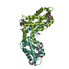
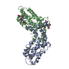
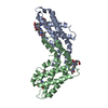
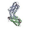
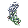
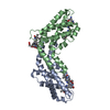
| Title | Crystal structure of c-phycocyanin of synechococcus vulcanus at 1.6 angstroms | |||||||||
 Components Components |
| |||||||||
 Keywords Keywords | PHOTOSYNTHESIS / CYANOBACTERIA / PHOTOSYSTEM II / LIGHT HARVESTING PROTEINS / THERMOSTABILITY | |||||||||
| Function / homology |  Function and homology information Function and homology informationphycobilisome / plasma membrane-derived thylakoid membrane / photosynthesis Similarity search - Function | |||||||||
| Biological species |  Thermosynechococcus vulcanus (bacteria) Thermosynechococcus vulcanus (bacteria) | |||||||||
| Method |  X-RAY DIFFRACTION / SYNCHROTRON / MOLECULAR REPLACEMENT / Resolution: 1.6 Å X-RAY DIFFRACTION / SYNCHROTRON / MOLECULAR REPLACEMENT / Resolution: 1.6 Å | |||||||||
 Authors Authors | Adir, N. / Dobrovetsky, E. / Lerner, N. | |||||||||
 Citation Citation | Journal: Biochim.Biophys.Acta / Year: 2002 Title: Refined structure of c-phycocyanin from the cyanobacterium Synechococcus vulcanus at 1.6 A: insights into the role of solvent molecules in thermal stability and co-factor structure Authors: Adir, N. / Vainer, R. / Lerner, N. | |||||||||
| History |
|
- Structure visualization
| Structure viewer | Molecule: MolmilJmol/JSmol |
|---|
- Downloads & links
-Download
| PDBx/mmCIF format | 1ktp.cif.gz | 88.7 KB | Display | PDBx/mmCIF format |
|---|---|---|---|---|
| PDB format | pdb1ktp.ent.gz | 66.4 KB | Display | PDB format |
| PDBx/mmJSON format | 1ktp.json.gz | Tree view | PDBx/mmJSON format | |
| Others |  Other downloads Other downloads |
-Validation report
| Arichive directory | https://data.pdbj.org/pub/pdb/validation_reports/kt/1ktpftp://data.pdbj.org/pub/pdb/validation_reports/kt/1ktp | HTTPS FTP |
|---|
-Related structure data
| Related structure data | 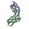 1i7yS S: Starting model for refinement |
|---|---|
| Similar structure data |


-Links
 PDBj
PDBj
- Assembly
| Deposited unit | 
| ||||||||
|---|---|---|---|---|---|---|---|---|---|
| 1 |
| ||||||||
| 2 | x 6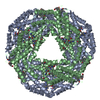
| ||||||||
| 3 | 
| ||||||||
| 4 | 
| ||||||||
| 5 | 
| ||||||||
| Unit cell |
|
-Components
| #1: Protein | Mass: 17470.656 Da / Num. of mol.: 1 Source method: isolated from a genetically manipulated source Source: (gene. exp.) Thermosynechococcus vulcanus (bacteria)Production host: | ||||
|---|---|---|---|---|---|
| #2: Protein | Mass: 18216.652 Da / Num. of mol.: 1 Source method: isolated from a genetically manipulated source Source: (gene. exp.) Thermosynechococcus vulcanus (bacteria)References: UniProt: P50033 | ||||
| #3: Chemical |   Mass: 588.694 Da / Num. of mol.: 3 / Source method: obtained synthetically / Formula: C33H40N4O6 Mass: 588.694 Da / Num. of mol.: 3 / Source method: obtained synthetically / Formula: C33H40N4O6#4: Water | ChemComp-HOH / |  Mass: 18.015 Da / Num. of mol.: 377 / Source method: isolated from a natural source / Formula: H2O Mass: 18.015 Da / Num. of mol.: 377 / Source method: isolated from a natural source / Formula: H2OHas protein modification | Y | |
-Experimental details
-Experiment
| Experiment | Method: X-RAY DIFFRACTION / Number of used crystals: 1 |
|---|
- Sample preparation
| Crystal | Density Matthews: 2.35 Å3/Da / Density % sol: 55.65 % | ||||||||||||||||||||
|---|---|---|---|---|---|---|---|---|---|---|---|---|---|---|---|---|---|---|---|---|---|
| Crystal grow | Temperature: 295 K / Method: vapor diffusion, hanging drop / pH: 7 Details: 5% PEG4000, 50MM HEPES, 5MG/ML PROTEIN, pH 7.00, VAPOR DIFFUSION, HANGING DROP, temperature 295K | ||||||||||||||||||||
| Crystal grow | *PLUS Temperature: 22 ℃ / pH: 8 / Details: Adir, N., (2001) J. Mol. Biol., 313, 71. | ||||||||||||||||||||
| Components of the solutions | *PLUS
|
-Data collection
| Diffraction | Mean temperature: 100 K |
|---|---|
| Diffraction source | Source: SYNCHROTRON / Site: CHESS  / Beamline: A1 / Wavelength: 0.928 / Beamline: A1 / Wavelength: 0.928 |
| Detector | Type: ADSC QUANTUM 4 / Detector: CCD / Date: Jun 16, 2001 |
| Radiation | Protocol: SINGLE WAVELENGTH / Monochromatic (M) / Laue (L): M / Scattering type: x-ray |
| Radiation wavelength | Wavelength: 0.928 Å / Relative weight: 1 |
| Reflection | Resolution: 1.6→20 Å / Num. all: 47823 / Num. obs: 47798 / % possible obs: 92.6 % / Observed criterion σ(I): -3 / Redundancy: 5.6 % / Biso Wilson estimate: 26.3 Å2 / Rmerge(I) obs: 0.079 / Net I/σ(I): 10 |
| Reflection shell | Resolution: 1.6→1.66 Å / Redundancy: 6.4 % / Rmerge(I) obs: 0.37 / Mean I/σ(I) obs: 3 / % possible all: 87.4 |
| Reflection | *PLUS Highest resolution: 1.6 Å / Lowest resolution: 20 Å / Num. obs: 51656 / % possible obs: 97.5 % / Redundancy: 5.6 % / Num. measured all: 419135 / Rmerge(I) obs: 0.078 |
| Reflection shell | *PLUS % possible obs: 99.3 % / Redundancy: 6.4 % / Rmerge(I) obs: 0.378 / Mean I/σ(I) obs: 3 |
- Processing
| Software |
| ||||||||||||||||||||||||||||||||||||||||||||||||||||||||||||
|---|---|---|---|---|---|---|---|---|---|---|---|---|---|---|---|---|---|---|---|---|---|---|---|---|---|---|---|---|---|---|---|---|---|---|---|---|---|---|---|---|---|---|---|---|---|---|---|---|---|---|---|---|---|---|---|---|---|---|---|---|---|
| Refinement | Method to determine structure: MOLECULAR REPLACEMENT Starting model: PDB ENTRY 1I7Y Resolution: 1.6→20 Å / Isotropic thermal model: ISOTROPIC / Cross valid method: THROUGHOUT / σ(F): 3 / Stereochemistry target values: ENGH & HUBER
| ||||||||||||||||||||||||||||||||||||||||||||||||||||||||||||
| Displacement parameters | Biso mean: 21.7 Å2 | ||||||||||||||||||||||||||||||||||||||||||||||||||||||||||||
| Refine analyze | Luzzati coordinate error obs: 0.21 Å / Luzzati d res low obs: 5 Å / Luzzati sigma a obs: 0.17 Å | ||||||||||||||||||||||||||||||||||||||||||||||||||||||||||||
| Refinement step | Cycle: LAST / Resolution: 1.6→20 Å
| ||||||||||||||||||||||||||||||||||||||||||||||||||||||||||||
| Refine LS restraints |
| ||||||||||||||||||||||||||||||||||||||||||||||||||||||||||||
| LS refinement shell | Resolution: 1.6→1.66 Å / Total num. of bins used: 10
| ||||||||||||||||||||||||||||||||||||||||||||||||||||||||||||
| Software | *PLUS Name: CNS / Version: 1 / Classification: refinement | ||||||||||||||||||||||||||||||||||||||||||||||||||||||||||||
| Refinement | *PLUS Highest resolution: 1.6 Å / Lowest resolution: 20 Å / Num. reflection obs: 35632 / % reflection Rfree: 7.7 % / Rfactor obs: 0.212 / Rfactor Rfree: 0.254 | ||||||||||||||||||||||||||||||||||||||||||||||||||||||||||||
| Solvent computation | *PLUS | ||||||||||||||||||||||||||||||||||||||||||||||||||||||||||||
| Displacement parameters | *PLUS Biso mean: 22.6 Å2 | ||||||||||||||||||||||||||||||||||||||||||||||||||||||||||||
| Refine LS restraints | *PLUS
| ||||||||||||||||||||||||||||||||||||||||||||||||||||||||||||
| LS refinement shell | *PLUS Rfactor Rfree: 0.3195 / % reflection Rfree: 10.8 % / Rfactor Rwork: 0.28 / Num. reflection obs: 3977 / Rfactor obs: 0.302 |
