 Movie
Movie Controller

[English] 日本語
 Yorodumi
- PDB-1i7y: CRYSTAL STRUCTURE OF C-PHYCOCYANIN OF SYNECHOCOCCUS VULCANUS AT 2... -
+ Open data

- Basic information
| Entry | Database: PDB / ID: 1i7y | |||||||||
|---|---|---|---|---|---|---|---|---|---|---|
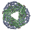
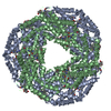
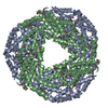
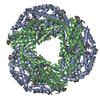
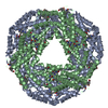
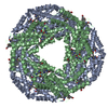
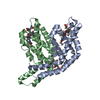
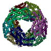
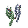
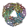
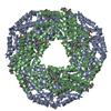
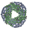
| Title | CRYSTAL STRUCTURE OF C-PHYCOCYANIN OF SYNECHOCOCCUS VULCANUS AT 2.5 ANGSTROMS. | |||||||||
 Components Components |
| |||||||||
 Keywords Keywords | PHOTOSYNTHESIS / Cyanobacteria / Photosystem II / Light harvesting proteins / thermostability | |||||||||
| Function / homology |  Function and homology information Function and homology informationphycobilisome / plasma membrane-derived thylakoid membrane / photosynthesis Similarity search - Function | |||||||||
| Biological species |  Thermostichus vulcanus (bacteria) Thermostichus vulcanus (bacteria) | |||||||||
| Method |  X-RAY DIFFRACTION / MOLECULAR REPLACEMENT / Resolution: 2.5 Å X-RAY DIFFRACTION / MOLECULAR REPLACEMENT / Resolution: 2.5 Å | |||||||||
 Authors Authors | Adir, N. / Dobrovetsky, E. / Lerner, N. | |||||||||
 Citation Citation | Journal: J.Mol.Biol. / Year: 2001 Title: Structure of c-phycocyanin from the thermophilic cyanobacterium Synechococcus vulcanus at 2.5 A: structural implications for thermal stability in phycobilisome assembly. Authors: Adir, N. / Dobrovetsky, Y. / Lerner, N. | |||||||||
| History |
|
- Structure visualization
| Structure viewer | Molecule: MolmilJmol/JSmol |
|---|
- Downloads & links
-Download
| PDBx/mmCIF format | 1i7y.cif.gz | 79.9 KB | Display | PDBx/mmCIF format |
|---|---|---|---|---|
| PDB format | pdb1i7y.ent.gz | 60.4 KB | Display | PDB format |
| PDBx/mmJSON format | 1i7y.json.gz | Tree view | PDBx/mmJSON format | |
| Others |  Other downloads Other downloads |
-Validation report
| Arichive directory | https://data.pdbj.org/pub/pdb/validation_reports/i7/1i7yftp://data.pdbj.org/pub/pdb/validation_reports/i7/1i7y | HTTPS FTP |
|---|
-Related structure data
| Related structure data |  1cbcS S: Starting model for refinement |
|---|---|
| Similar structure data |


-Links
 PDBj
PDBj
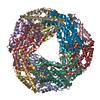
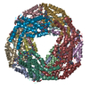
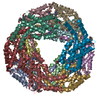
- Assembly
| Deposited unit | 
| ||||||||
|---|---|---|---|---|---|---|---|---|---|
| 1 | x 6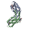
| ||||||||
| Unit cell |
|
-Components
| #1: Protein | Mass: 17470.656 Da / Num. of mol.: 1 / Source method: isolated from a natural source / Source: (natural) Thermostichus vulcanus (bacteria) / Genus: Thermosynechococcus / References: UniProt: Q9AM02 | ||
|---|---|---|---|
| #2: Protein | Mass: 18216.652 Da / Num. of mol.: 1 / Source method: isolated from a natural source / Source: (natural) Thermostichus vulcanus (bacteria) / Genus: Thermosynechococcus / References: UniProt: Q71RW8 | ||
| #3: Chemical |   Mass: 588.694 Da / Num. of mol.: 3 / Source method: obtained synthetically / Formula: C33H40N4O6 Mass: 588.694 Da / Num. of mol.: 3 / Source method: obtained synthetically / Formula: C33H40N4O6#4: Water | ChemComp-HOH / |  Mass: 18.015 Da / Num. of mol.: 88 / Source method: isolated from a natural source / Formula: H2O Mass: 18.015 Da / Num. of mol.: 88 / Source method: isolated from a natural source / Formula: H2O |
-Experimental details
-Experiment
| Experiment | Method: X-RAY DIFFRACTION / Number of used crystals: 2 |
|---|
- Sample preparation
| Crystal | Density Matthews: 2.74 Å3/Da / Density % sol: 58.07 % | ||||||||||||||||||||
|---|---|---|---|---|---|---|---|---|---|---|---|---|---|---|---|---|---|---|---|---|---|
| Crystal grow | Temperature: 290 K / Method: vapor diffusion, hanging drop / pH: 8 Details: 5% PEG4000, 50mM HEPES, 5mg/ml phycocyanin, pH 8.0, VAPOR DIFFUSION, HANGING DROP, temperature 290K | ||||||||||||||||||||
| Crystal grow | *PLUS Temperature: 22 ℃ | ||||||||||||||||||||
| Components of the solutions | *PLUS
|
-Data collection
| Diffraction | Mean temperature: 293 K |
|---|---|
| Diffraction source | Source: ROTATING ANODE / Type: RIGAKU RU200 / Wavelength: 1.5418 Å |
| Detector | Type: RIGAKU RAXIS IIC / Detector: IMAGE PLATE / Date: Sep 18, 2000 |
| Radiation | Monochromator: Graphite / Protocol: SINGLE WAVELENGTH / Monochromatic (M) / Laue (L): M / Scattering type: x-ray |
| Radiation wavelength | Wavelength: 1.5418 Å / Relative weight: 1 |
| Reflection | Resolution: 2.5→500 Å / Num. all: 195576 / Num. obs: 14116 / % possible obs: 97.5 % / Observed criterion σ(F): 0 / Observed criterion σ(I): 0 / Redundancy: 5.6 % / Biso Wilson estimate: 38.8 Å2 / Rmerge(I) obs: 0.09 / Net I/σ(I): 17.6 |
| Reflection shell | Resolution: 2.5→2.59 Å / Redundancy: 6.4 % / Rmerge(I) obs: 0.368 / Mean I/σ(I) obs: 4.8 / Num. unique all: 1429 / % possible all: 99.3 |
| Reflection | *PLUS Num. measured all: 112515 / Rmerge(I) obs: 0.09 |
| Reflection shell | *PLUS % possible obs: 99.3 % |
- Processing
| Software |
| |||||||||||||||||||||||||
|---|---|---|---|---|---|---|---|---|---|---|---|---|---|---|---|---|---|---|---|---|---|---|---|---|---|---|
| Refinement | Method to determine structure: MOLECULAR REPLACEMENT Starting model: PDB ENTRY 1CBC SUBUNITS A AND B Resolution: 2.5→500 Å / Isotropic thermal model: ISotropic / Cross valid method: THROUGHOUT / σ(F): 0 / σ(I): 0 / Stereochemistry target values: Engh & Huber
| |||||||||||||||||||||||||
| Displacement parameters | Biso mean: 26.4 Å2 | |||||||||||||||||||||||||
| Refine analyze |
| |||||||||||||||||||||||||
| Refinement step | Cycle: LAST / Resolution: 2.5→500 Å
| |||||||||||||||||||||||||
| Refine LS restraints |
| |||||||||||||||||||||||||
| LS refinement shell | Resolution: 2.5→2.59 Å / Total num. of bins used: 10
| |||||||||||||||||||||||||
| Software | *PLUS Name: CNS / Version: 1 / Classification: refinement | |||||||||||||||||||||||||
| Refinement | *PLUS Highest resolution: 2.5 Å / σ(F): 0 / % reflection Rfree: 9.5 % / Rfactor obs: 0.202 | |||||||||||||||||||||||||
| Solvent computation | *PLUS | |||||||||||||||||||||||||
| Displacement parameters | *PLUS Biso mean: 26.4 Å2 | |||||||||||||||||||||||||
| Refine LS restraints | *PLUS
| |||||||||||||||||||||||||
| LS refinement shell | *PLUS Highest resolution: 2.5 Å / Rfactor Rfree: 0.279 / % reflection Rfree: 11 % / Rfactor Rwork: 0.247 |
