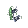
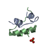
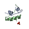
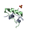
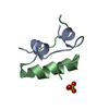
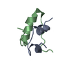
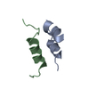
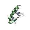
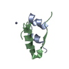
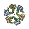
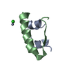
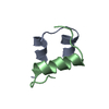
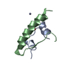
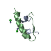
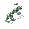
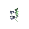
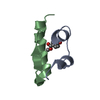
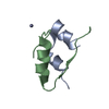
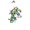
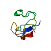
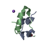
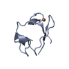
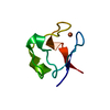
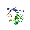
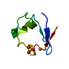
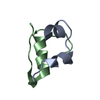
- PDB-4a7e: X-ray crystal structure of porcine insulin flash-cooled at high p... -
+
Open data
ID or keywords:
Loading...
-
Basic information
Entry
Database: PDB / ID: 4a7e
Title
X-ray crystal structure of porcine insulin flash-cooled at high pressure
Components
INSULIN A CHAIN
INSULIN B CHAIN
Keywords
HORMONE / HIGH-PRESSURE COOLING / HIGH-PRESSURE PROTEIN CRYSTALLOGRAPHY
Function / homology
Function and homology information
positive regulation of lipoprotein lipase activity / Insulin processing / IRS activation / Signal attenuation / Insulin receptor signalling cascade / Signaling by Insulin receptor / Synthesis, secretion, and deacylation of Ghrelin / PI5P, PP2A and IER3 Regulate PI3K/AKT Signaling / Insulin receptor recycling / response to L-arginine ...positive regulation of lipoprotein lipase activity / Insulin processing / IRS activation / Signal attenuation / Insulin receptor signalling cascade / Signaling by Insulin receptor / Synthesis, secretion, and deacylation of Ghrelin / PI5P, PP2A and IER3 Regulate PI3K/AKT Signaling / Insulin receptor recycling / response to L-arginine / lactate biosynthetic process / positive regulation of glucose metabolic process / lipoprotein biosynthetic process / positive regulation of fatty acid biosynthetic process / glycoprotein biosynthetic process / COPI-mediated anterograde transport / negative regulation of glycogen catabolic process / : / negative regulation of fatty acid metabolic process / negative regulation of feeding behavior / lipid biosynthetic process / negative regulation of acute inflammatory response / positive regulation of respiratory burst / TORC1 signaling / alpha-beta T cell activation / negative regulation of protein secretion / positive regulation of dendritic spine maintenance / negative regulation of gluconeogenesis / fatty acid homeostasis / positive regulation of glycogen biosynthetic process / positive regulation of insulin receptor signaling pathway / negative regulation of lipid catabolic process / negative regulation of respiratory burst involved in inflammatory response / negative regulation of ubiquitin-dependent protein catabolic process / nitric oxide-cGMP-mediated signaling / regulation of protein localization to plasma membrane / negative regulation of reactive oxygen species biosynthetic process / insulin-like growth factor receptor binding / neuron projection maintenance / positive regulation of mitotic nuclear division / positive regulation of glycolytic process / positive regulation of DNA replication / acute-phase response / positive regulation of cytokine production / positive regulation of D-glucose import across plasma membrane / insulin receptor binding / positive regulation of protein secretion / positive regulation of translation / wound healing / positive regulation of neuron projection development / hormone activity / negative regulation of protein catabolic process / positive regulation of protein localization to nucleus / glucose metabolic process / vasodilation / insulin receptor signaling pathway / glucose homeostasis / protease binding / positive regulation of MAPK cascade / positive regulation of canonical NF-kappaB signal transduction / positive regulation of phosphatidylinositol 3-kinase/protein kinase B signal transduction / positive regulation of cell migration / G protein-coupled receptor signaling pathway / negative regulation of gene expression / positive regulation of cell population proliferation / : / identical protein binding Similarity search - Function
Insulin / Insulin family / Insulin-like / Insulin/IGF/Relaxin family / Insulin / insulin-like growth factor / relaxin family. / Insulin, conserved site / Insulin family signature. / Insulin-like superfamily Similarity search - Domain/homology
In the structure databanks used in Yorodumi, some data are registered as the other names, "COVID-19 virus" and "2019-nCoV". Here are the details of the virus and the list of structure data.
Jan 31, 2019. EMDB accession codes are about to change! (news from PDBe EMDB page)
EMDB accession codes are about to change! (news from PDBe EMDB page)
The allocation of 4 digits for EMDB accession codes will soon come to an end. Whilst these codes will remain in use, new EMDB accession codes will include an additional digit and will expand incrementally as the available range of codes is exhausted. The current 4-digit format prefixed with “EMD-” (i.e. EMD-XXXX) will advance to a 5-digit format (i.e. EMD-XXXXX), and so on. It is currently estimated that the 4-digit codes will be depleted around Spring 2019, at which point the 5-digit format will come into force.
The EM Navigator/Yorodumi systems omit the EMD- prefix.
Related info.:Q: What is EMD? / ID/Accession-code notation in Yorodumi/EM Navigator
Yorodumi is a browser for structure data from EMDB, PDB, SASBDB, etc.
This page is also the successor to EM Navigator detail page, and also detail information page/front-end page for Omokage search.
The word "yorodu" (or yorozu) is an old Japanese word meaning "ten thousand". "mi" (miru) is to see.
Related info.:EMDB / PDB / SASBDB / Comparison of 3 databanks / Yorodumi Search / Aug 31, 2016. New EM Navigator & Yorodumi / Yorodumi Papers / Jmol/JSmol / Function and homology information / Changes in new EM Navigator and Yorodumi
 Movie
Movie Controller

 Yorodumi
 Open data
 Basic information
 Components
 Keywords
 Function and homology information
 X-RAY DIFFRACTION /
  Authors
 Citation
 Structure visualization
 Downloads & links
 Other downloads
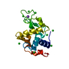


 PDBj
PDBj


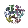
 Assembly
 Mass: 18.015 Da / Num. of mol.: 55 / Source method: isolated from a natural source / Formula: H2O
 Sample preparation
 / Beamline: X06DA / Wavelength: 0.9253
  Processing