 Movie
Movie Controller


+ Open data

- Basic information
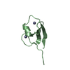
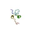
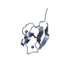
| Entry | Database: PDB / ID: 6khz | ||||||
|---|---|---|---|---|---|---|---|
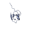
| Title | p62/SQSTM1 ZZ domain with Gly-peptide | ||||||
 Components Components | Sequestosome-1 | ||||||
 Keywords Keywords | SIGNALING PROTEIN / p62 | ||||||
| Function / homology |  Function and homology information Function and homology informationprotein localization to perinuclear region of cytoplasm / brown fat cell proliferation / protein targeting to vacuole involved in autophagy / regulation of Ras protein signal transduction / intracellular membraneless organelle / aggrephagy / negative regulation of toll-like receptor 4 signaling pathway / response to mitochondrial depolarisation / amphisome / regulation of protein complex stability ...protein localization to perinuclear region of cytoplasm / brown fat cell proliferation / protein targeting to vacuole involved in autophagy / regulation of Ras protein signal transduction / intracellular membraneless organelle / aggrephagy / negative regulation of toll-like receptor 4 signaling pathway / response to mitochondrial depolarisation / amphisome / regulation of protein complex stability / autophagy of mitochondrion / endosome organization / pexophagy / membraneless organelle assembly / regulation of mitochondrion organization / phagophore assembly site / ubiquitin-modified protein reader activity / regulation of canonical NF-kappaB signal transduction / Nuclear events mediated by NFE2L2 / aggresome / K63-linked polyubiquitin modification-dependent protein binding / endosomal transport / cellular response to stress / Lewy body / temperature homeostasis / negative regulation of ferroptosis / autolysosome / molecular sequestering activity / immune system process / mitophagy / energy homeostasis / inclusion body / signaling adaptor activity / negative regulation of protein ubiquitination / positive regulation of autophagy / ionotropic glutamate receptor binding / SH2 domain binding / autophagosome / p75NTR recruits signalling complexes / response to ischemia / NF-kB is activated and signals survival / Pexophagy / NRIF signals cell death from the nucleus / protein kinase C binding / protein catabolic process / positive regulation of long-term synaptic potentiation / positive regulation of protein localization to plasma membrane / PINK1-PRKN Mediated Mitophagy / sarcomere / macroautophagy / ubiquitin binding / protein sequestering activity / molecular condensate scaffold activity / P-body / receptor tyrosine kinase binding / PML body / autophagy / Interleukin-1 signaling / protein import into nucleus / Signaling by ALK fusions and activated point mutants / intracellular protein localization / late endosome / KEAP1-NFE2L2 pathway / signaling receptor activity / Neddylation / sperm midpiece / transcription by RNA polymerase II / ubiquitin-dependent protein catabolic process / protein-macromolecule adaptor activity / cell differentiation / intracellular signal transduction / positive regulation of apoptotic process / apoptotic process / ubiquitin protein ligase binding / protein kinase binding / protein-containing complex binding / glutamatergic synapse / enzyme binding / negative regulation of transcription by RNA polymerase II / endoplasmic reticulum / positive regulation of transcription by RNA polymerase II / mitochondrion / extracellular exosome / zinc ion binding / nucleoplasm / identical protein binding / cytoplasm / cytosol Similarity search - Function | ||||||
| Biological species |  Homo sapiens (human) Homo sapiens (human) | ||||||
| Method |  X-RAY DIFFRACTION / MOLECULAR REPLACEMENT / Resolution: 2.8 Å X-RAY DIFFRACTION / MOLECULAR REPLACEMENT / Resolution: 2.8 Å | ||||||
 Authors Authors | Kwon, D.H. / Kim, L. / Song, H.K. | ||||||
 Citation Citation | Journal: J.Biol.Chem. / Year: 2020 Title: Use of the LC3B-fusion technique for biochemical and structural studies of proteins involved in the N-degron pathway. Authors: Kim, L. / Kwon, D.H. / Heo, J. / Park, M.R. / Song, H.K. | ||||||
| History |
|
- Structure visualization
| Structure viewer | Molecule: MolmilJmol/JSmol |
|---|
- Downloads & links
-Download
| PDBx/mmCIF format | 6khz.cif.gz | 77.6 KB | Display | PDBx/mmCIF format |
|---|---|---|---|---|
| PDB format | pdb6khz.ent.gz | 59.8 KB | Display | PDB format |
| PDBx/mmJSON format | 6khz.json.gz | Tree view | PDBx/mmJSON format | |
| Others |  Other downloads Other downloads |
-Validation report
| Arichive directory | https://data.pdbj.org/pub/pdb/validation_reports/kh/6khzftp://data.pdbj.org/pub/pdb/validation_reports/kh/6khz | HTTPS FTP |
|---|
-Related structure data
| Related structure data | 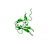 6kgiC 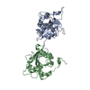 6kgjC 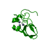 6lhnC  5yp7S S: Starting model for refinement C: citing same article ( |
|---|---|
| Similar structure data |


-Links
 PDBj
PDBj


- Assembly
| Deposited unit | 
| ||||||||
|---|---|---|---|---|---|---|---|---|---|
| 1 | 
| ||||||||
| 2 | 
| ||||||||
| 3 | 
| ||||||||
| 4 | 
| ||||||||
| Unit cell |
|
-Components
| #1: Protein/peptide | Mass: 5413.129 Da / Num. of mol.: 4 / Fragment: ZZ domain Source method: isolated from a genetically manipulated source Source: (gene. exp.) Homo sapiens (human) / Gene: SQSTM1, ORCA, OSIL / Production host:  #2: Chemical | ChemComp-ZN /   Mass: 65.409 Da / Num. of mol.: 8 / Source method: obtained synthetically / Formula: Zn Mass: 65.409 Da / Num. of mol.: 8 / Source method: obtained synthetically / Formula: ZnHas ligand of interest | N | Sequence details | The residues 121-125 GEEED is chimeric sequence. | |
|---|
-Experimental details
-Experiment
| Experiment | Method: X-RAY DIFFRACTION / Number of used crystals: 1 |
|---|
- Sample preparation
| Crystal | Density Matthews: 3.04 Å3/Da / Density % sol: 59.49 % |
|---|---|
| Crystal grow | Temperature: 293 K / Method: vapor diffusion, sitting drop / pH: 6.5 Details: 0.17 M Ammonium sulfate, 0.085 M Sodium cacodylate trihydrate pH 6.5, 22-30 % w/v Polyethylene glycol 8000 |
-Data collection
| Diffraction | Mean temperature: 173 K / Serial crystal experiment: N |
|---|---|
| Diffraction source | Source: ROTATING ANODE / Type: RIGAKU RU300 / Wavelength: 1 Å |
| Detector | Type: RIGAKU RAXIS IV / Detector: IMAGE PLATE / Date: Apr 14, 2017 |
| Radiation | Protocol: SINGLE WAVELENGTH / Monochromatic (M) / Laue (L): M / Scattering type: x-ray |
| Radiation wavelength | Wavelength: 1 Å / Relative weight: 1 |
| Reflection | Resolution: 2.8→50 Å / Num. obs: 6200 / % possible obs: 99.9 % / Redundancy: 6.7 % / Rmerge(I) obs: 0.13 / Rpim(I) all: 0.054 / Net I/σ(I): 19.8 |
| Reflection shell | Resolution: 2.8→2.85 Å / Rmerge(I) obs: 0.86 / Num. unique obs: 6200 |
- Processing
| Software |
| ||||||||||||||||
|---|---|---|---|---|---|---|---|---|---|---|---|---|---|---|---|---|---|
| Refinement | Method to determine structure: MOLECULAR REPLACEMENT Starting model: 5YP7 Resolution: 2.8→40.3 Å / Cross valid method: FREE R-VALUE
| ||||||||||||||||
| Refinement step | Cycle: LAST / Resolution: 2.8→40.3 Å
|
