 Movie
Movie Controller


+ Open data

- Basic information
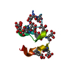
| Entry | Database: PDB / ID: 1dei | ||||||
|---|---|---|---|---|---|---|---|
| Title | DESHEPTAPEPTIDE (B24-B30) INSULIN | ||||||
 Components Components | (INSULIN) x 2 | ||||||
 Keywords Keywords | HORMONE / GLUCOSE METABOLISM | ||||||
| Function / homology |  Function and homology information Function and homology informationpositive regulation of lipoprotein lipase activity / Insulin processing / IRS activation / Signal attenuation / Insulin receptor signalling cascade / Signaling by Insulin receptor / Synthesis, secretion, and deacylation of Ghrelin / PI5P, PP2A and IER3 Regulate PI3K/AKT Signaling / Insulin receptor recycling / response to L-arginine ...positive regulation of lipoprotein lipase activity / Insulin processing / IRS activation / Signal attenuation / Insulin receptor signalling cascade / Signaling by Insulin receptor / Synthesis, secretion, and deacylation of Ghrelin / PI5P, PP2A and IER3 Regulate PI3K/AKT Signaling / Insulin receptor recycling / response to L-arginine / lactate biosynthetic process / positive regulation of glucose metabolic process / lipoprotein biosynthetic process / positive regulation of fatty acid biosynthetic process / glycoprotein biosynthetic process / COPI-mediated anterograde transport / negative regulation of glycogen catabolic process / : / negative regulation of fatty acid metabolic process / negative regulation of feeding behavior / lipid biosynthetic process / negative regulation of acute inflammatory response / positive regulation of respiratory burst / TORC1 signaling / alpha-beta T cell activation / negative regulation of protein secretion / positive regulation of dendritic spine maintenance / negative regulation of gluconeogenesis / fatty acid homeostasis / positive regulation of glycogen biosynthetic process / positive regulation of insulin receptor signaling pathway / negative regulation of lipid catabolic process / negative regulation of respiratory burst involved in inflammatory response / negative regulation of ubiquitin-dependent protein catabolic process / nitric oxide-cGMP-mediated signaling / regulation of protein localization to plasma membrane / negative regulation of reactive oxygen species biosynthetic process / insulin-like growth factor receptor binding / neuron projection maintenance / positive regulation of mitotic nuclear division / positive regulation of glycolytic process / positive regulation of DNA replication / acute-phase response / positive regulation of cytokine production / positive regulation of D-glucose import across plasma membrane / insulin receptor binding / positive regulation of protein secretion / positive regulation of translation / wound healing / positive regulation of neuron projection development / hormone activity / negative regulation of protein catabolic process / positive regulation of protein localization to nucleus / glucose metabolic process / vasodilation / insulin receptor signaling pathway / glucose homeostasis / protease binding / positive regulation of MAPK cascade / positive regulation of canonical NF-kappaB signal transduction / positive regulation of phosphatidylinositol 3-kinase/protein kinase B signal transduction / positive regulation of cell migration / G protein-coupled receptor signaling pathway / negative regulation of gene expression / positive regulation of cell population proliferation / : / identical protein binding Similarity search - Function | ||||||
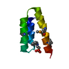
| Biological species |  | ||||||
| Method |  X-RAY DIFFRACTION / MOLECULAR REPLACEMENT / Resolution: 1.6 Å X-RAY DIFFRACTION / MOLECULAR REPLACEMENT / Resolution: 1.6 Å | ||||||
 Authors Authors | Bao, S.-J. / Chang, W.-R. / Wan, Z.-L. / Zhang, J.-P. / Liang, D.-C. | ||||||
 Citation Citation | Journal: Proc.Natl.Acad.Sci.USA / Year: 1997 Title: Crystal structure of desheptapeptide(B24-B30)insulin at 1.6 A resolution: implications for receptor binding. Authors: Bao, S.J. / Xie, D.L. / Zhang, J.P. / Chang, W.R. / Liang, D.C. #1: Journal: Sci.Sin., Ser.B (Engl.Ed.) / Year: 1987Title: Refinement of the Structure of Despentapeptide (B26-B30) Insulin at 1.5 Angstroms Resolution Authors: Dai, J.B. / Lou, M.Z. / You, J.M. / Liang, D.C. #2: Journal: Sci.Sin., Ser.B (Engl.Ed.) / Year: 1982Title: Crystallographic Studies on Desheptapeptide(B24-B30) Insulin-Growth of Single Crystals and Determination of its Crystallographic Parameters Authors: Chang, W.R. / Xie, D.L. / Liang, D.C. / al., et | ||||||
| History |
|
- Structure visualization
| Structure viewer | Molecule: MolmilJmol/JSmol |
|---|
- Downloads & links
-Download
| PDBx/mmCIF format | 1dei.cif.gz | 28.7 KB | Display | PDBx/mmCIF format |
|---|---|---|---|---|
| PDB format | pdb1dei.ent.gz | 19.8 KB | Display | PDB format |
| PDBx/mmJSON format | 1dei.json.gz | Tree view | PDBx/mmJSON format | |
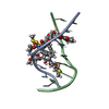
| Others |  Other downloads Other downloads |
-Validation report
| Arichive directory | https://data.pdbj.org/pub/pdb/validation_reports/de/1deiftp://data.pdbj.org/pub/pdb/validation_reports/de/1dei | HTTPS FTP |
|---|
-Related structure data
| Similar structure data |
|---|


-Links
 PDBj
PDBj


- Assembly
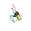
| Deposited unit | 
| ||||||||
|---|---|---|---|---|---|---|---|---|---|
| 1 | 
| ||||||||
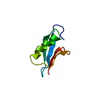
| 2 | 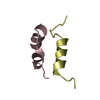
| ||||||||
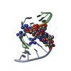
| 3 |
| ||||||||
| Unit cell |
| ||||||||
| Details | THE CRYSTALLOGRAPHIC ASYMMETRIC UNIT OF INSULIN CONSISTS OF TWO INSULIN MOLECULES EACH CONSISTING OF TWO CHAINS. THIS ENTRY PRESENTS COORDINATES FOR MOLECULES I (CHAIN IDENTIFIERS *A* AND *B*) AND II (CHAIN IDENTIFIERS *C* AND *D*). |
-Components
| #1: Protein/peptide | Mass: 2383.698 Da / Num. of mol.: 2 / Mutation: CHAIN B, DEL(24-30) Source method: isolated from a genetically manipulated source Details: DESHEPTAPEPTIDE (B24-B30) / Source: (gene. exp.) #2: Protein/peptide | Mass: 2547.929 Da / Num. of mol.: 2 / Mutation: CHAIN B, DEL(24-30) Source method: isolated from a genetically manipulated source Details: DESHEPTAPEPTIDE (B24-B30) / Source: (gene. exp.) #3: Water | ChemComp-HOH / |  Mass: 18.015 Da / Num. of mol.: 85 / Source method: isolated from a natural source / Formula: H2O Mass: 18.015 Da / Num. of mol.: 85 / Source method: isolated from a natural source / Formula: H2OHas protein modification | Y | |
|---|
-Experimental details
-Experiment
| Experiment | Method: X-RAY DIFFRACTION / Number of used crystals: 2 |
|---|
- Sample preparation
| Crystal | Density Matthews: 1.88 Å3/Da / Density % sol: 35 % |
|---|---|
| Crystal grow | *PLUS Method: other / Details: Chang, W.R., (1982) Sci. Sin. Ser. B, 25, 385. |
-Data collection
| Diffraction | Mean temperature: 293.1 K |
|---|---|
| Diffraction source | Source: ROTATING ANODE / Type: RIGAKU / Wavelength: 1.5418 |
| Detector | Type: STOE / Detector: DIFFRACTOMETER / Date: May 11, 1985 |
| Radiation | Monochromatic (M) / Laue (L): M / Scattering type: x-ray |
| Radiation wavelength | Wavelength: 1.5418 Å / Relative weight: 1 |
| Reflection | Resolution: 1.6→8 Å / Num. obs: 8331 / % possible obs: 81.1 % / Observed criterion σ(I): 1 / Redundancy: 2 % / Rmerge(I) obs: 0.088 |
| Reflection shell | Resolution: 1.6→1.63 Å / % possible all: 44.8 |
- Processing
| Software |
| ||||||||||||||||||||||||||||||||||||||||||||||||||||||||||||
|---|---|---|---|---|---|---|---|---|---|---|---|---|---|---|---|---|---|---|---|---|---|---|---|---|---|---|---|---|---|---|---|---|---|---|---|---|---|---|---|---|---|---|---|---|---|---|---|---|---|---|---|---|---|---|---|---|---|---|---|---|---|
| Refinement | Method to determine structure: MOLECULAR REPLACEMENT Starting model: DESPENTAPEPTIDE(B26-B30)INSULIN Resolution: 1.6→8 Å / σ(F): 1
| ||||||||||||||||||||||||||||||||||||||||||||||||||||||||||||
| Displacement parameters | Biso mean: 14.3 Å2 | ||||||||||||||||||||||||||||||||||||||||||||||||||||||||||||
| Refinement step | Cycle: LAST / Resolution: 1.6→8 Å
| ||||||||||||||||||||||||||||||||||||||||||||||||||||||||||||
| Refine LS restraints |
| ||||||||||||||||||||||||||||||||||||||||||||||||||||||||||||
| LS refinement shell | Resolution: 1.6→1.63 Å
| ||||||||||||||||||||||||||||||||||||||||||||||||||||||||||||
| Software | *PLUS Name: X-PLOR / Classification: refinement | ||||||||||||||||||||||||||||||||||||||||||||||||||||||||||||
| Refinement | *PLUS σ(F): 2 | ||||||||||||||||||||||||||||||||||||||||||||||||||||||||||||
| Solvent computation | *PLUS | ||||||||||||||||||||||||||||||||||||||||||||||||||||||||||||
| Displacement parameters | *PLUS | ||||||||||||||||||||||||||||||||||||||||||||||||||||||||||||
| Refine LS restraints | *PLUS
| ||||||||||||||||||||||||||||||||||||||||||||||||||||||||||||
| LS refinement shell | *PLUS Rfactor Rwork: 0.26 |
