 ムービー
ムービー コントローラー


+ データを開く

- 基本情報
基本情報
| 登録情報 | データベース: PDB / ID: 6nl6 | ||||||
|---|---|---|---|---|---|---|---|
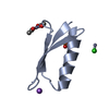
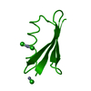
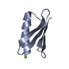
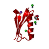
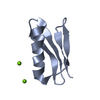
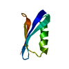
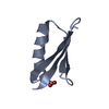
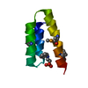
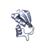
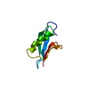
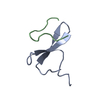
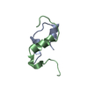
| タイトル | Crystal structure of mutant B1 immunoglobulin-binding domain of Streptococcal Protein G (T16F, T18A, V21E, T25L, K28Y, V29I, K31R, Q32H, Y33L, N35K, D36H, N37Q) | ||||||
 要素 要素 | Immunoglobulin G-binding protein G | ||||||
 キーワード キーワード | DE NOVO PROTEIN / Metal-mediated complex / B1 Domain of Streptococcal protein G / Immunoglobulin binding protein | ||||||
| 機能・相同性 |  機能・相同性情報 機能・相同性情報 | ||||||
| 生物種 |  Streptococcus (バクテリア) Streptococcus (バクテリア) | ||||||
| 手法 |  X線回折 / シンクロトロン / 分子置換 / 解像度: 1.4 Å X線回折 / シンクロトロン / 分子置換 / 解像度: 1.4 Å | ||||||
 データ登録者 データ登録者 | Huxford, T. / Stec, B. / Maniaci, B. | ||||||
 引用 引用 | ジャーナル: Biochemistry / 年: 2019 タイトル: Design of High-Affinity Metal-Controlled Protein Dimers. 著者: Maniaci, B. / Lipper, C.H. / Anipindi, D.L. / Erlandsen, H. / Cole, J.L. / Stec, B. / Huxford, T. / Love, J.J. | ||||||
| 履歴 |
|
- 構造の表示
構造の表示
| 構造ビューア | 分子: MolmilJmol/JSmol |
|---|
- ダウンロードとリンク
-ダウンロード
| PDBx/mmCIF形式 | 6nl6.cif.gz | 123.4 KB | 表示 | PDBx/mmCIF形式 |
|---|---|---|---|---|
| PDB形式 | pdb6nl6.ent.gz | 94.9 KB | 表示 | PDB形式 |
| PDBx/mmJSON形式 | 6nl6.json.gz | ツリー表示 | PDBx/mmJSON形式 | |
| その他 |  その他のダウンロード その他のダウンロード |
-検証レポート
| アーカイブディレクトリ | https://data.pdbj.org/pub/pdb/validation_reports/nl/6nl6ftp://data.pdbj.org/pub/pdb/validation_reports/nl/6nl6 | HTTPS FTP |
|---|
-関連構造データ


-リンク
 PDBj
PDBj


- 集合体
集合体
| 登録構造単位 | 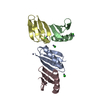
| ||||||||
|---|---|---|---|---|---|---|---|---|---|
| 1 | 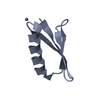
| ||||||||
| 2 | 
| ||||||||
| 3 | 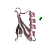
| ||||||||
| 4 | 
| ||||||||
| 単位格子 |
|
-要素
| #1: 抗体 | 分子量: 6348.063 Da / 分子数: 4 変異: T16F, T18A, V21E, T25L, K28Y, V29I, K31R, Q32H, Y33L, D36H, N35K, D36A, N37Q 由来タイプ: 組換発現 / 由来: (組換発現) Streptococcus (バクテリア) / 遺伝子: spg / プラスミド: pET21a / 詳細 (発現宿主): T7 expression / 発現宿主: #2: 化合物 | ChemComp-ZN /   分子量: 65.409 Da / 分子数: 6 / 由来タイプ: 合成 / 式: Zn / タイプ: SUBJECT OF INVESTIGATION 分子量: 65.409 Da / 分子数: 6 / 由来タイプ: 合成 / 式: Zn / タイプ: SUBJECT OF INVESTIGATION#3: 化合物 | ChemComp-CL /   分子量: 35.453 Da / 分子数: 4 / 由来タイプ: 合成 / 式: Cl 分子量: 35.453 Da / 分子数: 4 / 由来タイプ: 合成 / 式: Cl#4: 水 | ChemComp-HOH / |  分子量: 18.015 Da / 分子数: 264 / 由来タイプ: 天然 / 式: H2O 分子量: 18.015 Da / 分子数: 264 / 由来タイプ: 天然 / 式: H2O |
|---|
-実験情報
-実験
| 実験 | 手法: X線回折 / 使用した結晶の数: 1 |
|---|
- 試料調製
試料調製
| 結晶 | マシュー密度: 2.21 Å3/Da / 溶媒含有率: 44.33 % |
|---|---|
| 結晶化 | 温度: 298 K / 手法: 蒸気拡散法, ハンギングドロップ法 詳細: 70 mM acetic Acid pH 3.6, 30 mM acetic acid pH 5.8, 30% 2,4-methylpentanediol, 100 mM NaCl, and 20 mM zinc sulfate PH範囲: 3.6 - 5.8 |
-データ収集
| 回折 | 平均測定温度: 100 K / Serial crystal experiment: N | |||||||||||||||
|---|---|---|---|---|---|---|---|---|---|---|---|---|---|---|---|---|
| 放射光源 | 由来: シンクロトロン / サイト: APS  / ビームライン: 24-ID-C / 波長: 1.00002 Å / ビームライン: 24-ID-C / 波長: 1.00002 Å | |||||||||||||||
| 検出器 | タイプ: DECTRIS PILATUS 6M-F / 検出器: PIXEL / 日付: 2012年6月15日 | |||||||||||||||
| 放射 | プロトコル: SINGLE WAVELENGTH / 単色(M)・ラウエ(L): M / 散乱光タイプ: x-ray | |||||||||||||||
| 放射波長 | 波長: 1.00002 Å / 相対比: 1 | |||||||||||||||
| Reflection twin |
| |||||||||||||||
| 反射 | 解像度: 1.4→37.38 Å / Num. obs: 42380 / % possible obs: 95.7 % / 冗長度: 9.9 % / Rmerge(I) obs: 0.1 / Net I/σ(I): 7.8 | |||||||||||||||
| 反射 シェル | 解像度: 1.4→1.44 Å / 冗長度: 6.5 % / Rmerge(I) obs: 0.29 / Num. unique obs: 2092 / % possible all: 70.44 |
- 解析
解析
| ソフトウェア |
| ||||||||||||||||||||||||||||||||||||||||||||||||||||||||||||||||||||||||||||||||||||||||||||||||||||||||||||||||||||||||||||||||||||||||||||||||||||||||||||||||||||||||||||||||||||||
|---|---|---|---|---|---|---|---|---|---|---|---|---|---|---|---|---|---|---|---|---|---|---|---|---|---|---|---|---|---|---|---|---|---|---|---|---|---|---|---|---|---|---|---|---|---|---|---|---|---|---|---|---|---|---|---|---|---|---|---|---|---|---|---|---|---|---|---|---|---|---|---|---|---|---|---|---|---|---|---|---|---|---|---|---|---|---|---|---|---|---|---|---|---|---|---|---|---|---|---|---|---|---|---|---|---|---|---|---|---|---|---|---|---|---|---|---|---|---|---|---|---|---|---|---|---|---|---|---|---|---|---|---|---|---|---|---|---|---|---|---|---|---|---|---|---|---|---|---|---|---|---|---|---|---|---|---|---|---|---|---|---|---|---|---|---|---|---|---|---|---|---|---|---|---|---|---|---|---|---|---|---|---|---|
| 精密化 | 構造決定の手法: 分子置換 開始モデル: 1PGA 解像度: 1.4→37.38 Å / Cor.coef. Fo:Fc: 0.981 / Cor.coef. Fo:Fc free: 0.962 / SU B: 2.131 / SU ML: 0.04 / 交差検証法: THROUGHOUT / ESU R: 0.015 / ESU R Free: 0.015 / 詳細: HYDROGENS HAVE BEEN ADDED IN THE RIDING POSITIONS
| ||||||||||||||||||||||||||||||||||||||||||||||||||||||||||||||||||||||||||||||||||||||||||||||||||||||||||||||||||||||||||||||||||||||||||||||||||||||||||||||||||||||||||||||||||||||
| 溶媒の処理 | イオンプローブ半径: 0.8 Å / 減衰半径: 0.8 Å / VDWプローブ半径: 1.2 Å | ||||||||||||||||||||||||||||||||||||||||||||||||||||||||||||||||||||||||||||||||||||||||||||||||||||||||||||||||||||||||||||||||||||||||||||||||||||||||||||||||||||||||||||||||||||||
| 原子変位パラメータ | Biso mean: 17.271 Å2
| ||||||||||||||||||||||||||||||||||||||||||||||||||||||||||||||||||||||||||||||||||||||||||||||||||||||||||||||||||||||||||||||||||||||||||||||||||||||||||||||||||||||||||||||||||||||
| 精密化ステップ | サイクル: 1 / 解像度: 1.4→37.38 Å
| ||||||||||||||||||||||||||||||||||||||||||||||||||||||||||||||||||||||||||||||||||||||||||||||||||||||||||||||||||||||||||||||||||||||||||||||||||||||||||||||||||||||||||||||||||||||
| 拘束条件 |
|