 Movie
Movie Controller

[English] 日本語
 Yorodumi
- PDB-3fil: Structural and energetic determinants for hyperstable variants of... -
+ Open data

- Basic information
| Entry | Database: PDB / ID: 3fil | ||||||
|---|---|---|---|---|---|---|---|
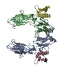
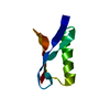
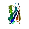
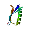
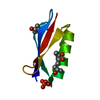
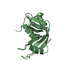
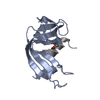
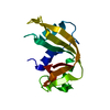
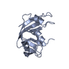
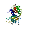
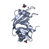
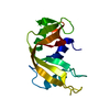
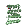
| Title | Structural and energetic determinants for hyperstable variants of GB1 obtained from in-vitro evolution | ||||||
 Components Components | (Immunoglobulin G-binding protein G) x 2 | ||||||
 Keywords Keywords | PROTEIN BINDING / dimerization / beta sheet / alpha helix / improved hydrophobic packing of core residues / Cell wall / IgG-binding protein / Peptidoglycan-anchor / Secreted | ||||||
| Function / homology |  Function and homology information Function and homology information | ||||||
| Biological species |  Streptococcus sp. 'group G' (bacteria) Streptococcus sp. 'group G' (bacteria) | ||||||
| Method |  X-RAY DIFFRACTION / SYNCHROTRON / MOLECULAR REPLACEMENT / Resolution: 0.88 Å X-RAY DIFFRACTION / SYNCHROTRON / MOLECULAR REPLACEMENT / Resolution: 0.88 Å | ||||||
 Authors Authors | Max, K.E.A. / Heinemann, U. | ||||||
 Citation Citation | Journal: J.Mol.Biol. / Year: 2009 Title: Dimer Formation of a Stabilized Gbeta1 Variant: A Structural and Energetic Analysis Authors: Thoms, S. / Max, K.E.A. / Wunderlich, M. / Jacso, T. / Lilie, H. / Reif, B. / Heinemann, U. / Schmid, F.X. | ||||||
| History |
|
- Structure visualization
| Structure viewer | Molecule: MolmilJmol/JSmol |
|---|
- Downloads & links
-Download
| PDBx/mmCIF format | 3fil.cif.gz | 70.3 KB | Display | PDBx/mmCIF format |
|---|---|---|---|---|
| PDB format | pdb3fil.ent.gz | 52.1 KB | Display | PDB format |
| PDBx/mmJSON format | 3fil.json.gz | Tree view | PDBx/mmJSON format | |
| Others |  Other downloads Other downloads |
-Validation report
| Arichive directory | https://data.pdbj.org/pub/pdb/validation_reports/fi/3filftp://data.pdbj.org/pub/pdb/validation_reports/fi/3fil | HTTPS FTP |
|---|
-Related structure data
| Related structure data | 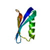 1pgaS S: Starting model for refinement |
|---|---|
| Similar structure data |


-Links
 PDBj
PDBj

- Assembly
| Deposited unit | 
| ||||||||
|---|---|---|---|---|---|---|---|---|---|
| 1 |
| ||||||||
| Unit cell |
|
-Components
| #1: Antibody | Mass: 6249.999 Da / Num. of mol.: 1 Fragment: immunoglobulin binding domain, UNP residues 303-357 Mutation: T2Q, E15V, T16L, T18I, N37L Source method: isolated from a genetically manipulated source Source: (gene. exp.) Streptococcus sp. 'group G' (bacteria) / Strain: G148 / Gene: spg / Plasmid: pET11 / Production host: |
|---|---|
| #2: Antibody | Mass: 6221.989 Da / Num. of mol.: 1 Fragment: immunoglobulin binding domain, UNP residues 303-357 Mutation: T2Q, E15V, T16L, T18I, N37L Source method: isolated from a genetically manipulated source Source: (gene. exp.) Streptococcus sp. 'group G' (bacteria) / Strain: G148 / Gene: spg / Plasmid: pET11 / Production host: |
| #3: Chemical | ChemComp-CA /   Mass: 40.078 Da / Num. of mol.: 1 / Source method: obtained synthetically / Formula: Ca Mass: 40.078 Da / Num. of mol.: 1 / Source method: obtained synthetically / Formula: Ca |
| #4: Water | ChemComp-HOH /  Mass: 18.015 Da / Num. of mol.: 239 / Source method: isolated from a natural source / Formula: H2O Mass: 18.015 Da / Num. of mol.: 239 / Source method: isolated from a natural source / Formula: H2O |
| Has protein modification | Y |
| Sequence details | FME IS INITIATING N-FORMYLMETHIONINE THAT IS THE BIOLOGICAL STARTING RESIDUE IN E. COLI WHICH HAS ...FME IS INITIATING |
-Experimental details
-Experiment
| Experiment | Method: X-RAY DIFFRACTION / Number of used crystals: 1 |
|---|
- Sample preparation
| Crystal | Density Matthews: 1.93 Å3/Da / Density % sol: 39.4 % |
|---|---|
| Crystal grow | Temperature: 293.15 K / Method: vapor diffusion, hanging drop / pH: 5.6 Details: protein solution: 50mM sodium acetate pH 5.6, 50mg/ml protein. reservoir solution: 25% PEG 3350, 0.1M citric acids pH 3.5. Drop 400nl protein solution & 400nl reservoir solution, 80 micro-l ...Details: protein solution: 50mM sodium acetate pH 5.6, 50mg/ml protein. reservoir solution: 25% PEG 3350, 0.1M citric acids pH 3.5. Drop 400nl protein solution & 400nl reservoir solution, 80 micro-l reservoir, VAPOR DIFFUSION, HANGING DROP, temperature 293.15K |
-Data collection
| Diffraction | Mean temperature: 110 K |
|---|---|
| Diffraction source | Source: SYNCHROTRON / Site: BESSY  / Beamline: 14.2 / Wavelength: 0.88561 Å / Beamline: 14.2 / Wavelength: 0.88561 Å |
| Detector | Type: RAYONIX MX-225 / Detector: CCD / Date: Mar 6, 2006 Details: Mirror 1: Silicon, active surface 50nm Rh-coated. Mirror 2: Glas, active surface 50 nm Rh-coated. |
| Radiation | Monochromator: silicon-111 crystal / Protocol: SINGLE WAVELENGTH / Monochromatic (M) / Laue (L): M / Scattering type: x-ray |
| Radiation wavelength | Wavelength: 0.88561 Å / Relative weight: 1 |
| Reflection | Resolution: 0.842→19.466 Å / Num. all: 87156 / Num. obs: 86548 / % possible obs: 97.3 % / Observed criterion σ(F): 0 / Redundancy: 7.08 % / Biso Wilson estimate: 11.564 Å2 / Rmerge(I) obs: 0.074 / Rsym value: 0.063 / Net I/σ(I): 13.26 |
| Reflection shell | Resolution: 0.84→0.98 Å / Redundancy: 5.82 % / Rmerge(I) obs: 0.481 / Mean I/σ(I) obs: 2.56 / Num. unique all: 9687 / Rsym value: 0.775 / % possible all: 84.8 |
- Processing
| Software |
| |||||||||||||||||||||||||||||||||
|---|---|---|---|---|---|---|---|---|---|---|---|---|---|---|---|---|---|---|---|---|---|---|---|---|---|---|---|---|---|---|---|---|---|---|
| Refinement | Method to determine structure: MOLECULAR REPLACEMENT Starting model: PDB ENTRY 1PGA Resolution: 0.88→19.466 Å / Num. parameters: 10668 / Num. restraintsaints: 13229 / Cross valid method: FREE R / σ(F): 0 / Stereochemistry target values: Engh & Huber Details: According to PROCHECK and WHATCHECK, no backbone outliers were reported. The ASN A8 backbone and sidechain groups are centered in their 2FOFC density
| |||||||||||||||||||||||||||||||||
| Refine analyze | Num. disordered residues: 17 / Occupancy sum hydrogen: 853 / Occupancy sum non hydrogen: 1102.25 | |||||||||||||||||||||||||||||||||
| Refinement step | Cycle: LAST / Resolution: 0.88→19.466 Å
| |||||||||||||||||||||||||||||||||
| Refine LS restraints |
|
